Copyright
©2011 Baishideng Publishing Group Co.
World J Stem Cells. Aug 26, 2011; 3(8): 70-82
Published online Aug 26, 2011. doi: 10.4252/wjsc.v3.i8.70
Published online Aug 26, 2011. doi: 10.4252/wjsc.v3.i8.70
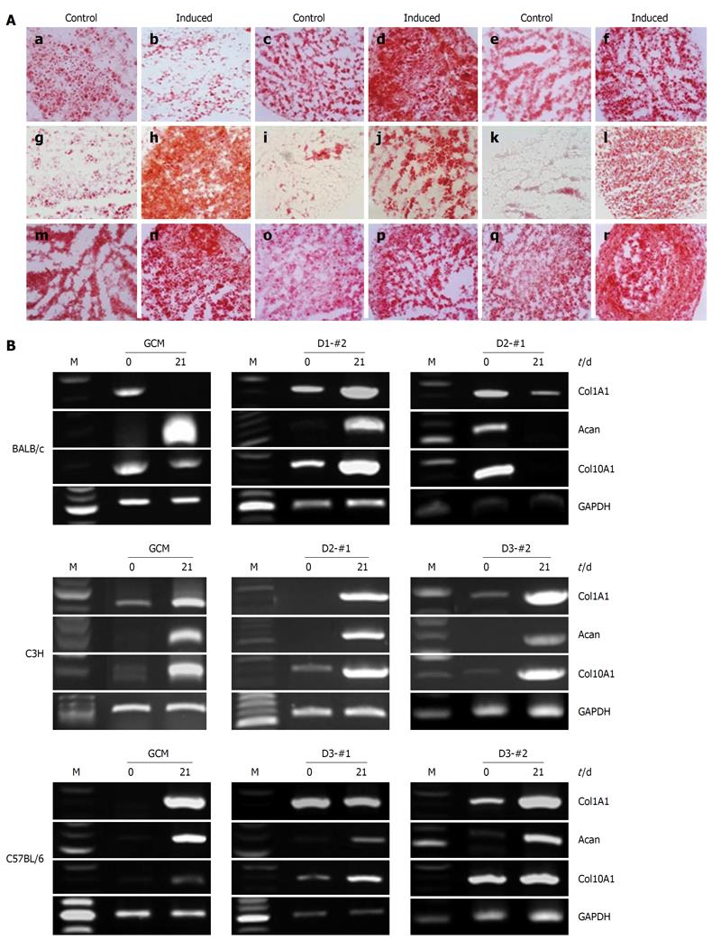
Figure 6 Chondrogenic differentiation potential of the established mouse clonal mesenchymal stem cell lines and nonclonal mesenchymal stem cells.
A: Histochemical staining with Safranin-O showed chondrogenically differentiated mouse clonal mesenchymal stem cell lines, tested 21 d after chondrogenic induction: a and b: BALB/c gradient centrifugation method (GCM); c and d: BALB/c D1-#2; e and f: BALB/c D2-#1; g and h: C3H GCM; i and j: C3H D2-#1; k and l: C3H D3-#2; m and n: C57BL/6 GCM; o and p: C57BL/6 D3-#1; q and r: C57BL/6 D3-#2; B: Reverse transcriptase-polymerase chain reaction analysis of chondrogenic markers, type II collagen, aggrecan, and type X collagen at days 0 and 21 after chondrogenic induction. Glyceraldehyde phosphate dehydrogenase was used as an internal control.
- Citation: Jeon MS, Yi TG, Lim HJ, Moon SH, Lee MH, Kang JS, Kim CS, Lee DH, Song SU. Characterization of mouse clonal mesenchymal stem cell lines established by subfractionation culturing method. World J Stem Cells 2011; 3(8): 70-82
- URL: https://www.wjgnet.com/1948-0210/full/v3/i8/70.htm
- DOI: https://dx.doi.org/10.4252/wjsc.v3.i8.70


