Copyright
©2011 Baishideng Publishing Group Co.
World J Stem Cells. Aug 26, 2011; 3(8): 70-82
Published online Aug 26, 2011. doi: 10.4252/wjsc.v3.i8.70
Published online Aug 26, 2011. doi: 10.4252/wjsc.v3.i8.70
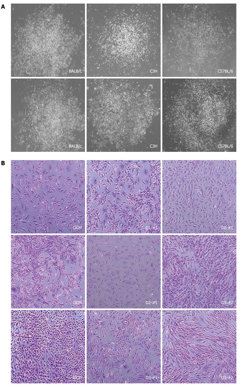
Figure 2 Pictures of the individual colonies and cell morphology of the established mouse clonal mesenchymal stem cell lines.
A: Representative pictures of single colonies obtained from different strains; B: Pictures of mouse clonal mesenchymal stem cells (mcMSCs) established from single colonies at about 70%-80% confluence. Nonclonal MSCs isolated by the conventional method, labeled as the gradient centrifugation method, from the three strains are also shown. Every mcMSCs in the right panel have a fibroblast-like morphology. The cells were fixed with 4% paraformaldehyde for 10 min and stained with 0.1% crystal violet for 5 min. Magnification: 100 ×.
- Citation: Jeon MS, Yi TG, Lim HJ, Moon SH, Lee MH, Kang JS, Kim CS, Lee DH, Song SU. Characterization of mouse clonal mesenchymal stem cell lines established by subfractionation culturing method. World J Stem Cells 2011; 3(8): 70-82
- URL: https://www.wjgnet.com/1948-0210/full/v3/i8/70.htm
- DOI: https://dx.doi.org/10.4252/wjsc.v3.i8.70


