Copyright
©2011 Baishideng Publishing Group Co.
World J Stem Cells. Apr 26, 2011; 3(4): 34-42
Published online Apr 26, 2011. doi: 10.4252/wjsc.v3.i4.34
Published online Apr 26, 2011. doi: 10.4252/wjsc.v3.i4.34
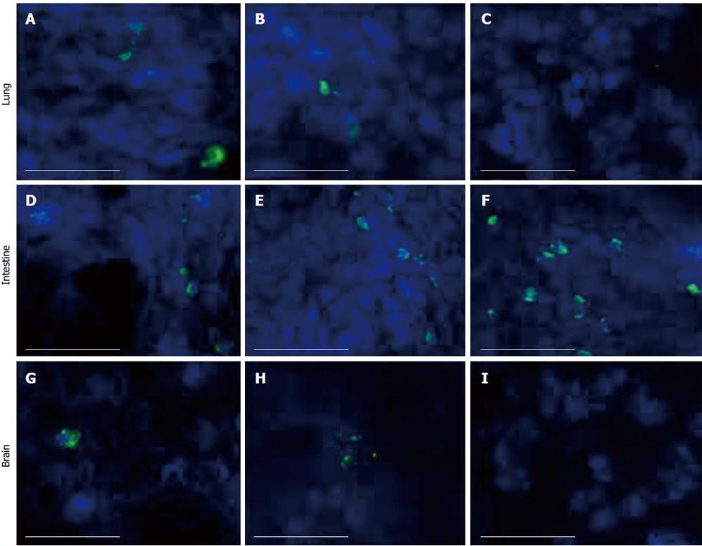
Figure 5 Detection of human umbilical cord matrix stem cells by immunofluorescence staining on days 1, 3 and 7.
Human umbilical cord matrix stem cells were detected in lung (A-C), intestine (D-F) and brain (G-I) using anti-human mitochondrial antibody. DAPI was used as counter stain for nuclear staining. All images were captured using 40 × objective. Scale bar = 50 μm.
- Citation: Maurya DK, Doi C, Pyle M, Rachakatla RS, Davis D, Tamura M, Troyer D. Non-random tissue distribution of human naïve umbilical cord matrix stem cells. World J Stem Cells 2011; 3(4): 34-42
- URL: https://www.wjgnet.com/1948-0210/full/v3/i4/34.htm
- DOI: https://dx.doi.org/10.4252/wjsc.v3.i4.34


