Copyright
©The Author(s) 2025.
World J Stem Cells. Apr 26, 2025; 17(4): 103919
Published online Apr 26, 2025. doi: 10.4252/wjsc.v17.i4.103919
Published online Apr 26, 2025. doi: 10.4252/wjsc.v17.i4.103919
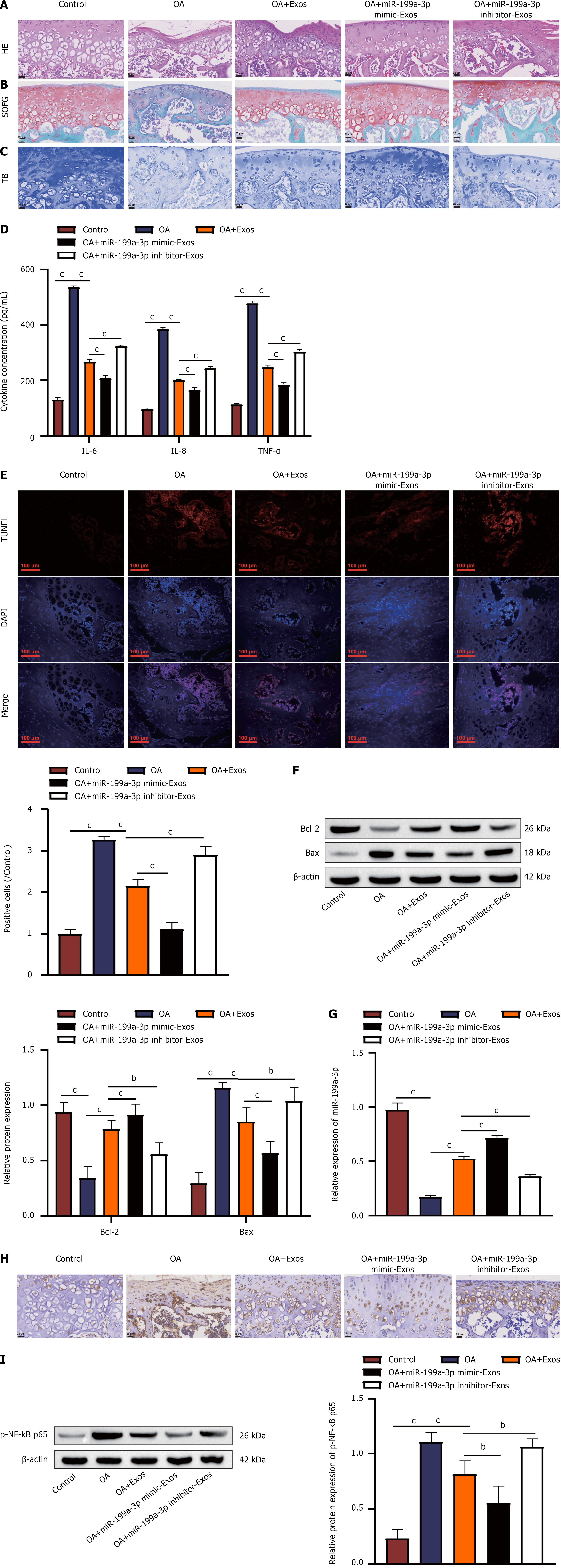
Figure 6 Human umbilical cord mesenchymal stem cell-derived exosomes relieve osteoarthritis progression in vivo through the delivery of miR-199a-3p.
A: Hematoxylin and eosin staining was used to examine the pathological conditions of rat articular cartilage tissues (scale bar = 25 μm); B: Safranin O/fast green staining was used to assess the quantity of chondrocytes within rat articular cartilage tissues (scale bar = 25 μm); C: Examination of cartilage differentiation in rat articular cartilage tissues via toluidine blue staining (scale bar = 25 μm); D: Interleukin-6, interleukin-8, and tumor necrosis factor-α levels in rat articular cartilage tissue were detected via enzyme-linked immunosorbent assay; E: Evaluation of cell apoptosis in rat articular cartilage tissue via terminal deoxynucleotidyl transferase-mediated deoxyuridine triphosphate-nick end labelling staining (scale bar = 100 μm); F: Western blot analysis was used to measure the expression of Bcl-2 and Bax apoptosis-related proteins; G: Quantitative real-time polymerase chain reaction was used to quantify miR-199a-3p expression in rat cartilage tissues; H: Immunohistochemistry was used to determine the presence of mitogen-activated protein kinase 4 in rat articular cartilage tissues (scale bar = 25 μm); I: P-nuclear factor-kappaB p65 expression in rat articular cartilage tissues was evaluated through western blot analysis. bP < 0.01, cP < 0.001. OA: Osteoarthritis; Exos: Exosomes; HE: Hematoxylin and eosin; TB: Toluidine blue; SOFG: Safranin O/fast green; NF-κB: Nuclear factor-kappaB; IL: Interleukin; TNF: Tumor necrosis factor; DAPI: 4’,6-diamidino-2-phenylindole; TUNEL: Terminal deoxynucleotidyl transferase-mediated deoxyuridine triphosphate-nick end labelling.
- Citation: Chen LQ, Ma S, Yu J, Zuo DC, Yin ZJ, Li FY, He X, Peng HT, Shi XQ, Huang WJ, Li Q, Wang J. Human umbilical cord mesenchymal stem cell-derived exosomal miR-199a-3p inhibits the MAPK4/NF-κB signaling pathway to relieve osteoarthritis. World J Stem Cells 2025; 17(4): 103919
- URL: https://www.wjgnet.com/1948-0210/full/v17/i4/103919.htm
- DOI: https://dx.doi.org/10.4252/wjsc.v17.i4.103919


