Copyright
©The Author(s) 2025.
World J Stem Cells. Feb 26, 2025; 17(2): 101395
Published online Feb 26, 2025. doi: 10.4252/wjsc.v17.i2.101395
Published online Feb 26, 2025. doi: 10.4252/wjsc.v17.i2.101395
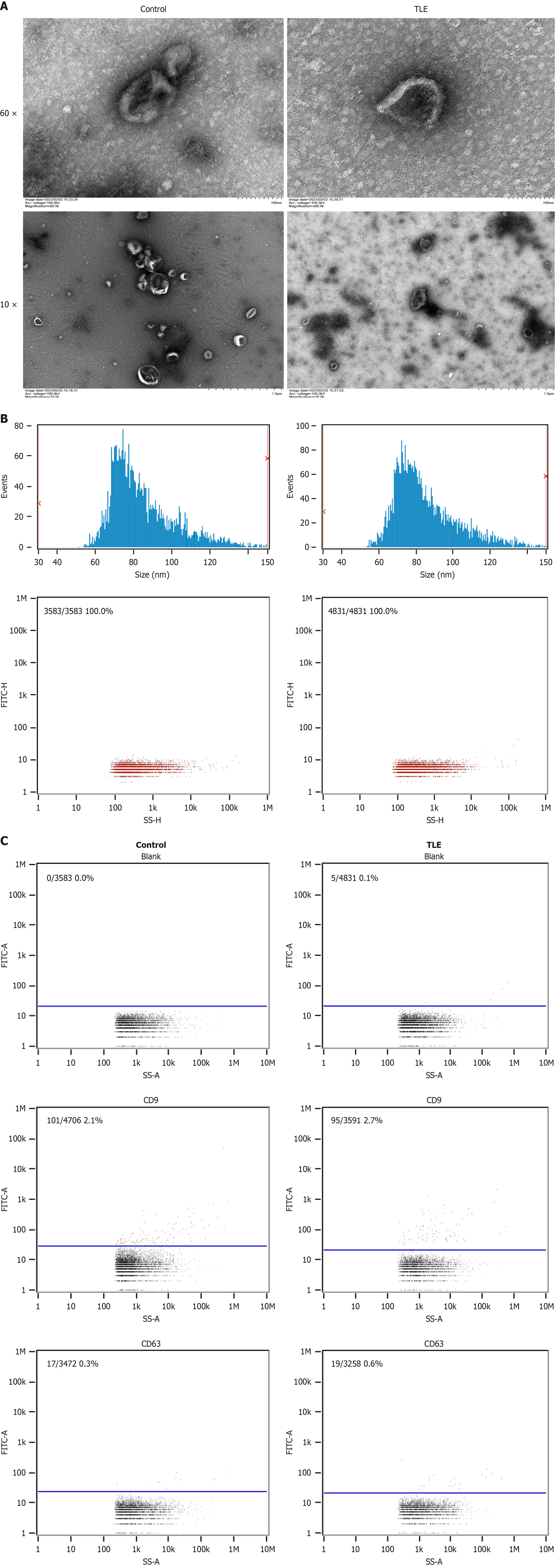
Figure 1 Characteristic of exosomes from bone marrow mesenchymal stem cells.
A: Transmission electron microscopy images reveal the characteristic “cup-shaped” morphology and clear membrane structure of exosomes from both the epilepsy and normal groups of mice; B: Nanoparticle tracking analysis shows the average particle size and concentration to be 8531 nm and 1.62E+10 particles/mL for the epilepsy group, and 84.64 nm and 3.29E+10 particles/mL for the normal group, respectively; C: Flow cytometry analysis demonstrates positive expression of the protein markers CD9 and CD63 in exosomes from both groups, with a slightly higher positive rate for CD9 in the epilepsy group than in the normal group and minimal expression of CD63 in both groups. TLE: Temporal lobe epilepsy.
- Citation: Wang W, Yin J. Exosomal miR-203 from bone marrow stem cells targets the SOCS3/NF-κB pathway to regulate neuroinflammation in temporal lobe epilepsy. World J Stem Cells 2025; 17(2): 101395
- URL: https://www.wjgnet.com/1948-0210/full/v17/i2/101395.htm
- DOI: https://dx.doi.org/10.4252/wjsc.v17.i2.101395


