Copyright
©The Author(s) 2025.
World J Stem Cells. Feb 26, 2025; 17(2): 101030
Published online Feb 26, 2025. doi: 10.4252/wjsc.v17.i2.101030
Published online Feb 26, 2025. doi: 10.4252/wjsc.v17.i2.101030
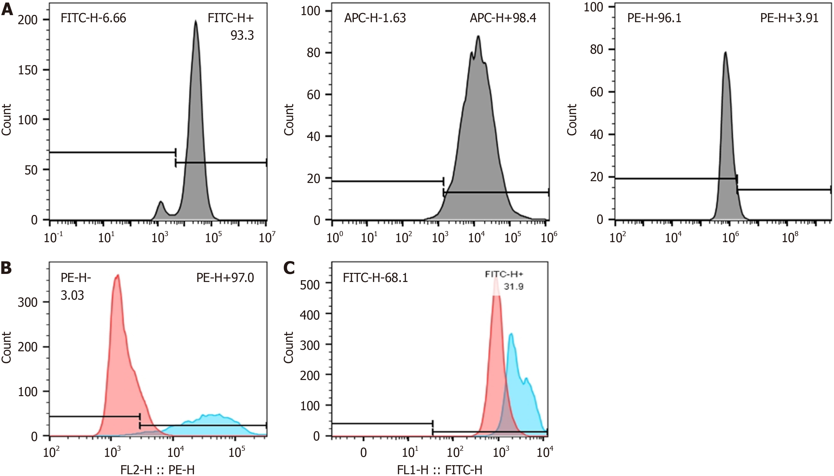
Figure 1 Characterization of fetal dermal mesenchymal stem cell M1/2 macrophages.
A: Detection of CD29 (FITC), CD44 (APC), and CD45 (PE) expression, characteristic markers of mesenchymal stem cells by flow cytometry; B: Flow cytometric analysis of CD86 (PE) cell surface marker expression (red - RAW264.7, blue - M1-type macrophages induced); C: Flow cytometric analysis of CD206 (FITC) cell surface marker expression (red - RAW264.7, blue - M2-type macrophages induced).
- Citation: Xia ZY, Wang Y, Shi N, Lu MQ, Deng YX, Qi YJ, Wang XL, Zhao J, Jiang DY. Fetal mice dermal mesenchymal stem cells promote wound healing by inducing M2 type macrophage polarization. World J Stem Cells 2025; 17(2): 101030
- URL: https://www.wjgnet.com/1948-0210/full/v17/i2/101030.htm
- DOI: https://dx.doi.org/10.4252/wjsc.v17.i2.101030


