Copyright
©The Author(s) 2025.
World J Stem Cells. Jan 26, 2025; 17(1): 101485
Published online Jan 26, 2025. doi: 10.4252/wjsc.v17.i1.101485
Published online Jan 26, 2025. doi: 10.4252/wjsc.v17.i1.101485
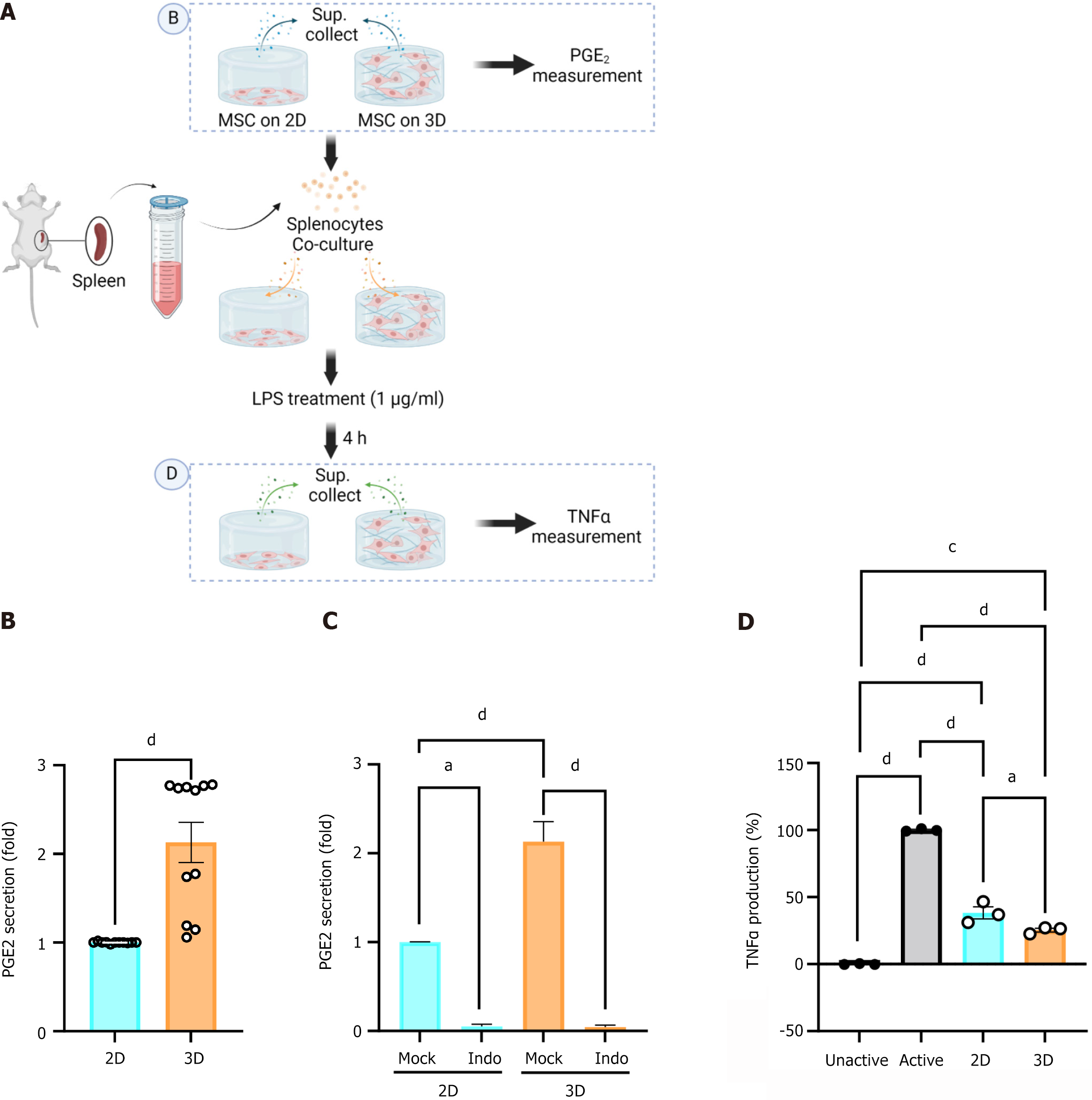
Figure 3 Three-dimensional-cultured mesenchymal stromal cells elevate the secretion of prostaglandin E2 and suppress tumor necrosis factor-alpha from lipopolysaccharide-stimulated splenocytes.
A: Schematic of the method for collection of supernatant from mesenchymal stromal cells (MSCs) or co-cultured MSCs and lipopolysaccharide-stimulated murine splenocytes; B: Secreted prostaglandin E2 (PGE2) in a MSC culture medium was determined by enzyme linked immunosorbent assay (ELISA) (n = 4 independent experiments, unpaired t-test, dP < 0.0001); C: MSCs co-cultured with splenocytes were treated with 10 mmol/L indomethacin (marked as Indo) and secreted PGE2 was assessed using ELISA (n = 3 independent experiment, one-way ANOVA, aP < 0.05, dP < 0.0001); D: Secreted tumor necrosis factor alpha from MSCs co-cultured with murine splenocytes was assessed by ELISA (n = 3 independent experiment, one-way ANOVA, aP < 0.05, cP < 0.001, dP < 0.0001). 2D: Two-dimensional; 3D: Three-dimensional; PGE2: Prostaglandin E2; LPS: Lipopolysaccharide; TNFα: Tumor necrosis factor alpha.
- Citation: Kim OH, Kang H, Chang ES, Lim Y, Seo YJ, Lee HJ. Extended protective effects of three dimensional cultured human mesenchymal stromal cells in a neuroinflammation model. World J Stem Cells 2025; 17(1): 101485
- URL: https://www.wjgnet.com/1948-0210/full/v17/i1/101485.htm
- DOI: https://dx.doi.org/10.4252/wjsc.v17.i1.101485


