Copyright
©The Author(s) 2024.
World J Stem Cells. Aug 26, 2024; 16(8): 811-823
Published online Aug 26, 2024. doi: 10.4252/wjsc.v16.i8.811
Published online Aug 26, 2024. doi: 10.4252/wjsc.v16.i8.811
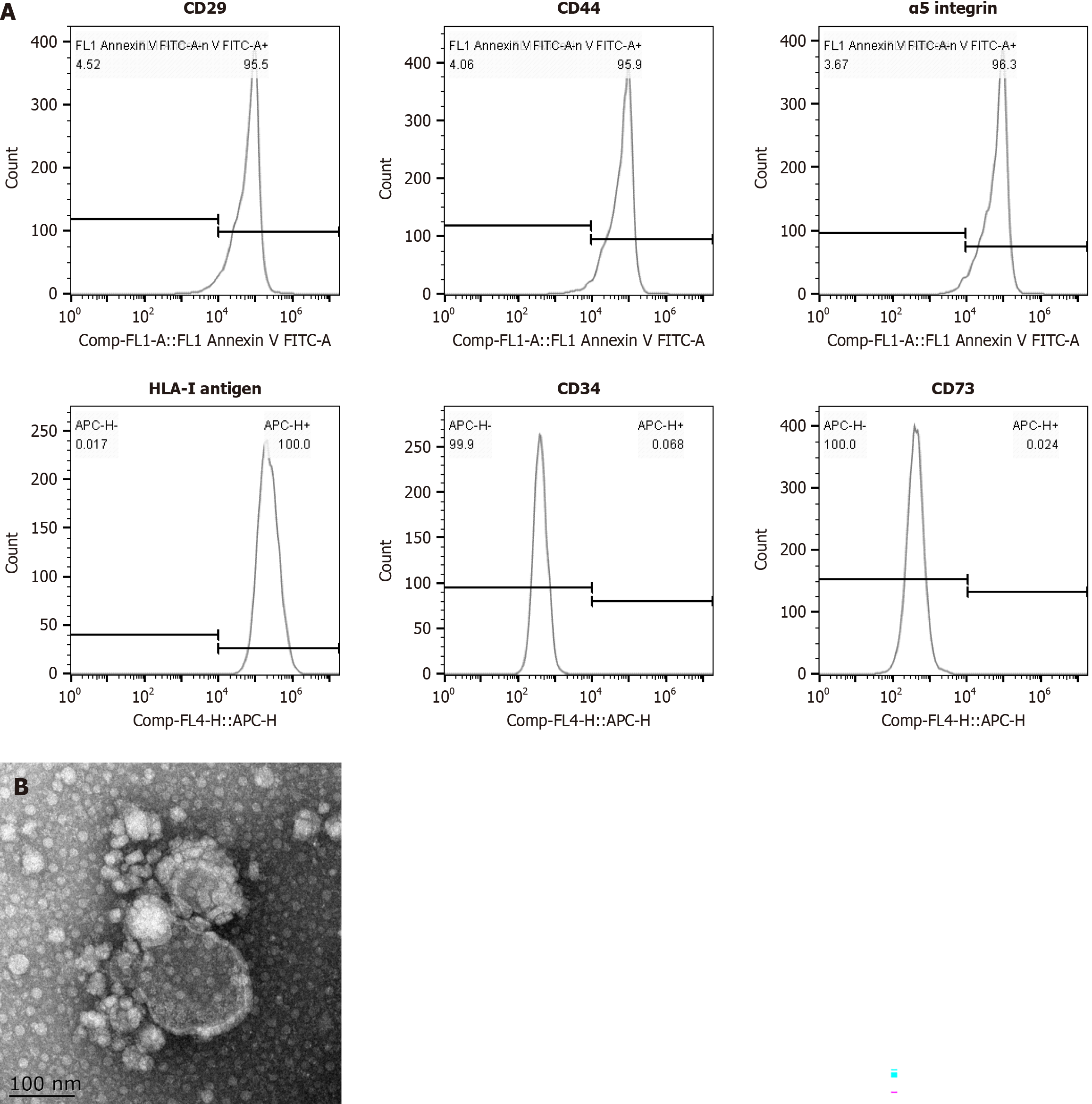
Figure 1 Detection of mesenchymal stromal cell-derived microvesicle markers by flow cytometry and transmission and scanning electron microscopy.
A: The expression of surface molecules (CD29, CD44, α5 integrins and the HLA-I antigen) was positive, whereas the expression of CD34 and CD73 was negative; B: Transmission and scanning electron microscopy were performed on purified mesenchymal stromal cell-derived microvesicles to reveal their spheroid morphologies and confirm their sizes.
- Citation: Chen QH, Zhang Y, Gu X, Yang PL, Yuan J, Yu LN, Chen JM. Microvesicles derived from mesenchymal stem cells inhibit acute respiratory distress syndrome-related pulmonary fibrosis in mouse partly through hepatocyte growth factor. World J Stem Cells 2024; 16(8): 811-823
- URL: https://www.wjgnet.com/1948-0210/full/v16/i8/811.htm
- DOI: https://dx.doi.org/10.4252/wjsc.v16.i8.811


