Copyright
©The Author(s) 2024.
World J Stem Cells. Jun 26, 2024; 16(6): 728-738
Published online Jun 26, 2024. doi: 10.4252/wjsc.v16.i6.728
Published online Jun 26, 2024. doi: 10.4252/wjsc.v16.i6.728
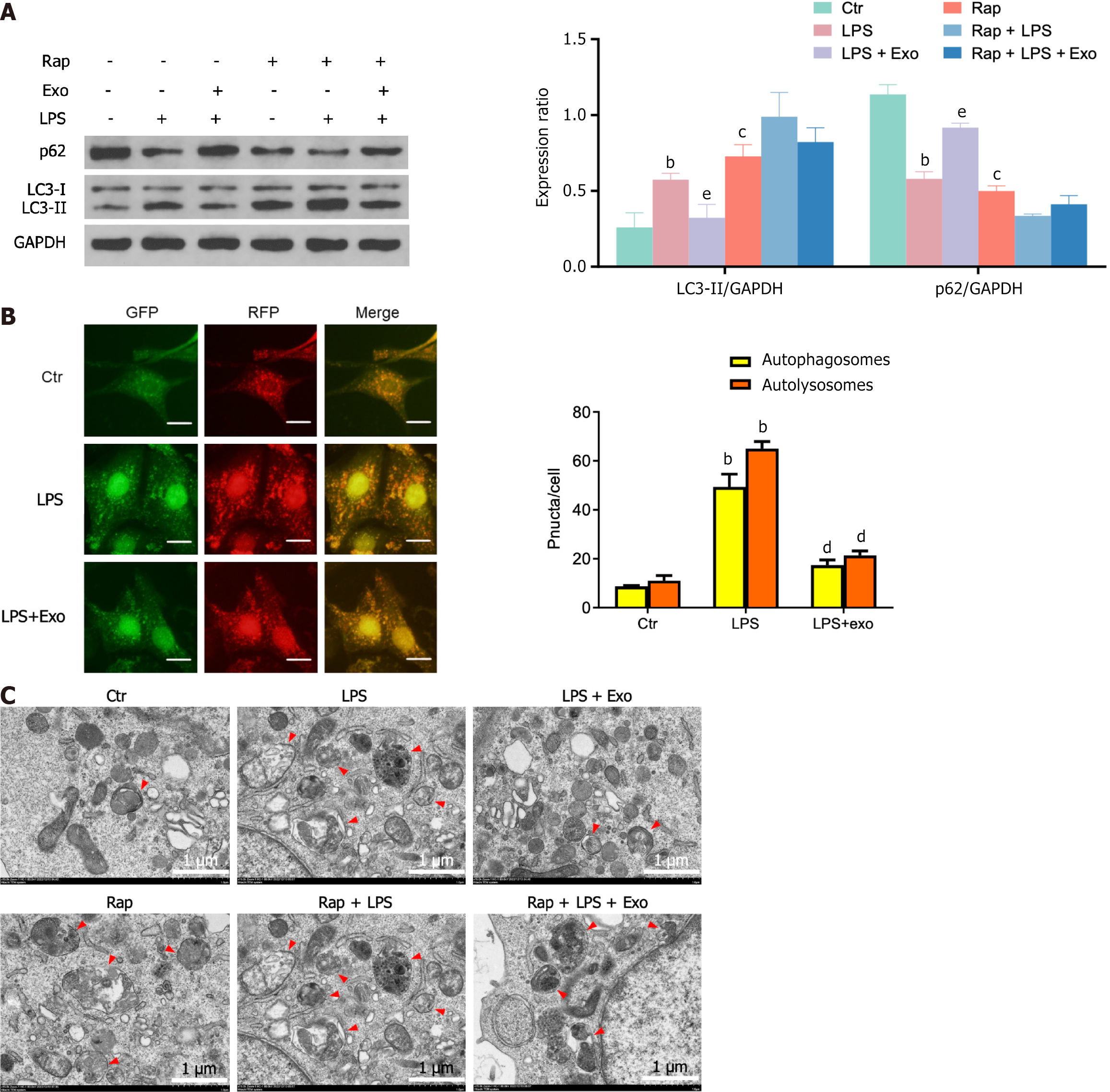
Figure 2 Human umbilical cord mesenchymal stem cell exosomes attenuate lipopolysaccharide-induced autophagy in intestinal epithelial cells.
A: The expression of p62 and LC3 in each cell group was determined by western blotting; B: IEC-18 cells were infected with Ad-mRFP-GFP-LC3 for 48 h and photographed with the confocal microscopy. Representative mRFP-LC3, GFP-LC3 and merge images were shown. The number of GFP-LC3 dots and mRFP-LC3 dots per cell were counted to quantify the number of autophagosomes (yellow dots) and autolysosomes (red dots) per cell; C: The ultrastructure of IEC-18 cells was presented by transmission electron microscopy, with red arrows indicating degradative autophagic vacuoles. The scale bar represents 1 μm. Data was expressed as mean ± SEM of three or six independent experiments. bP < 0.01 vs control group, cP < 0.001 vs control group; dP < 0.05 vs lipopolysaccharide, eP < 0.01 vs lipopolysaccharide. LPS: Lipopolysaccharide.
- Citation: Zhu L, He L, Duan W, Yang B, Li N. Umbilical cord mesenchymal stem cell exosomes alleviate necrotizing enterocolitis in neonatal mice by regulating intestinal epithelial cells autophagy. World J Stem Cells 2024; 16(6): 728-738
- URL: https://www.wjgnet.com/1948-0210/full/v16/i6/728.htm
- DOI: https://dx.doi.org/10.4252/wjsc.v16.i6.728


