Copyright
©The Author(s) 2024.
World J Stem Cells. Jun 26, 2024; 16(6): 728-738
Published online Jun 26, 2024. doi: 10.4252/wjsc.v16.i6.728
Published online Jun 26, 2024. doi: 10.4252/wjsc.v16.i6.728
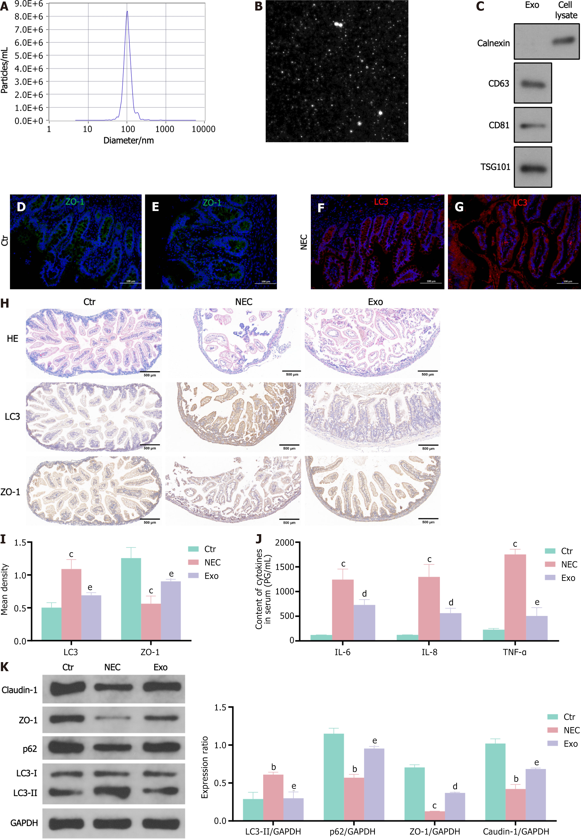
Figure 1 Intestinal epithelial cells autophagy increases and tight junction protein expression decreases in necrotizing enterocolitis neonates.
Human umbilical cord mesenchymal stem cell exosomes reduce autophagy and increase tight junction protein expression in the small intestine tissue of necrotizing enterocolitis mice. A-G: Exosomes particle size analysis (nanoparticle tracking analysis method) analyzes the size of human umbilical cord mesenchymal stem cell exosomes (hUCMSCs-exos) vesicles (A); morphology of isolated exosomes using transmission electron microscopy (B); expression levels of TSG101, CD81, CD63, and calnexin in hUCMSCs-exos were detected by western blot (C); expression of the intestinal epithelial cell-specific protein ZO-1 in neonatal colon tissue (green fluorescence) (D and E); expression of the autophagy protein LC3 in intestinal epithelial cells in neonatal colon tissue (red fluorescence), scale bar, 100 μm (F and G); H and I: Hematoxylin-eosin and immunohistochemical staining of LC3 and p62 in mouse distal ileum. Scale bar = 500 μm; J: Inflammatory factor levels in mice serum was detected by enzyme-linked immunosorbent assay; K: Western blot detects autophagy (LC3, p62) and tight junction (ZO-1, claudin-1) protein expression in mouse distal ileum tissue. Data was expressed as mean ± SEM of three or six independent experiments. bP < 0.01 vs control group, cP < 0.001 vs control group, dP < 0.05 vs necrotizing enterocolitis, eP < 0.01 vs necrotizing enterocolitis. NEC: Necrotizing enterocolitis; Ctr: Control; IL: Interleukin; TNF: Tumor necrosis factor.
- Citation: Zhu L, He L, Duan W, Yang B, Li N. Umbilical cord mesenchymal stem cell exosomes alleviate necrotizing enterocolitis in neonatal mice by regulating intestinal epithelial cells autophagy. World J Stem Cells 2024; 16(6): 728-738
- URL: https://www.wjgnet.com/1948-0210/full/v16/i6/728.htm
- DOI: https://dx.doi.org/10.4252/wjsc.v16.i6.728


