Copyright
©The Author(s) 2024.
World J Stem Cells. Jun 26, 2024; 16(6): 708-727
Published online Jun 26, 2024. doi: 10.4252/wjsc.v16.i6.708
Published online Jun 26, 2024. doi: 10.4252/wjsc.v16.i6.708
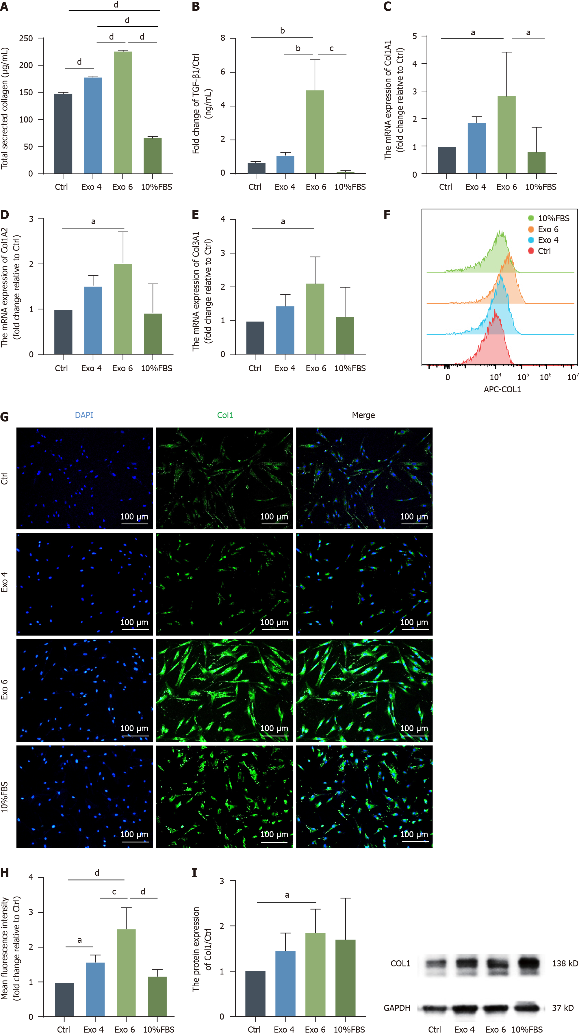
Figure 6 Human umbilical cord mesenchymal stromal cell-derived exosome promote the production of collagen in primary vaginal fibroblasts.
A: A Sirius Red collagen staining kit was used to determine the total secreted collagen level in the fibroblasts’ supernatant; B: Enzyme-linked immunosorbent assays were conducted to quantify the expression level of transforming growth factor-β1 in the cell culture supernatant; C-E: Quantitative real-time polymerase chain reaction was performed to evaluate the mRNA levels of Col1A1, Col1A2 and Col3A1; F-H: Representative histogram overlay showing the median fluorescence intensity of each group (F) and quantification of these values (H), the expression level of Col1 was also detected by immunofluorescence. Green indicates Col1; blue indicates DAPI-stained nuclei. Scale bar: 100 μm (G); I: The protein expression of Col1 was determined by western blot, and the results were quantified, which was in line with the previous results of Sirius Red staining, real-time polymerase chain reaction, FCM and immunofluorescence. The data are presented as the means ± SEMs. Statistical analysis was performed with one-way ANOVA. aP < 0.05; bP < 0.01; cP < 0.001; dP < 0.0001. FBS: Fetal bovine serum; TGF: Transforming growth factor; Exo: Exosome.
- Citation: Xu LM, Yu XX, Zhang N, Chen YS. Exosomes from umbilical cord mesenchymal stromal cells promote the collagen production of fibroblasts from pelvic organ prolapse. World J Stem Cells 2024; 16(6): 708-727
- URL: https://www.wjgnet.com/1948-0210/full/v16/i6/708.htm
- DOI: https://dx.doi.org/10.4252/wjsc.v16.i6.708


