Copyright
©The Author(s) 2024.
World J Stem Cells. May 26, 2024; 16(5): 575-590
Published online May 26, 2024. doi: 10.4252/wjsc.v16.i5.575
Published online May 26, 2024. doi: 10.4252/wjsc.v16.i5.575
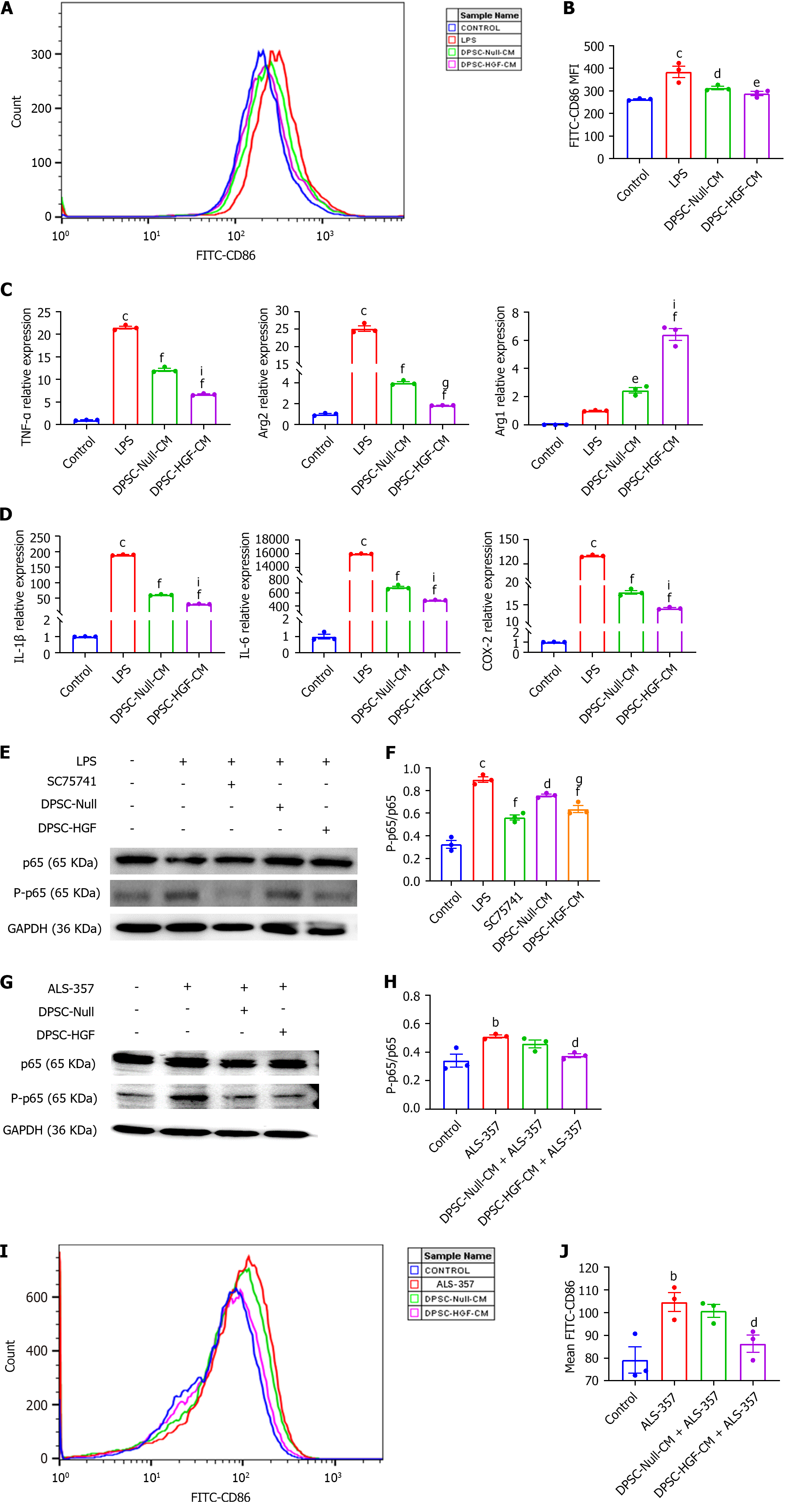
Figure 5 Effects of the supernatant of Ad-Null and Ad-hepatocyte growth factor modified dental pulp stem cells on the polarization and inflammatory cytokine expression of RAW264.
7 mouse macrophages under inflammatory stimulation. RAW264.7 cells were indirectly co-cultured with the supernatant of Ad-Null modified dental pulp stem cells (DPSCs) and Ad-hepatocyte growth factor modified DPSCs for 24 h under lipopolysaccharide (150 ng/mL) stimulation. The mean fluorescence intensity of the anti-CD86 antibody was detected by flow cytometry. A: Representative image; B: A statistical plot of the mean fluorescence intensity (MFI) of CD86 was generated (n = 3); C: The mRNA expression levels of the M1 macrophage markers tumor necrosis factor-α (TNF-α) and arginase 2 (Arg2) and the M2 macrophage marker Arg1 were detected by real-time reverse transcription polymerase chain reaction (RT-PCR) under inflammatory stimulation (n = 3); D: The mRNA expression levels of the inflammatory factors interleukin-1β (IL-1β), IL-6, and cyclooxygenase-2 were detected by RT-PCR under inflammatory stimulation (n = 3); E: The expression of phosphorylated p65 and p65 in RAW264.7 cells under inflammatory stimulation conditions was investigated using western blotting; F: The gray values were analyzed; G: The expression levels of phosphorylated p65 and p65 in RAW264.7 cells under nuclear factor-κB (NF-κB) activator treatment were detected by western blotting; H: Analyzed for gray values; I: After NF-κB activator treatment, the CD86 MFI was detected by flow cytometry; J: A statistical plot of the MFI of CD86 was generated (n = 3). All results are representative of the mean ± SEM; bP < 0.01, cP < 0.001 vs. control group; dP < 0.05, eP < 0.01, fP < 0.001 vs. lipopolysaccharide group; gP < 0.05, iP < 0.001 vs. supernatant of Ad-Null modified dental pulp stem cells group. LPS: Lipopolysaccharide; DPSC-Null-CM: Supernatant of Ad-Null modified dental pulp stem cells; DPSC-HGF-CM: Supernatant of Ad-hepatocyte growth factor modified dental pulp stem cells; IL: Interleukin; TNF: Tumor necrosis factor; Arg2: Arginase 2; COX-2: Cyclooxygenase-2; GAPDH: glyceraldehyde-3-phosphate dehydrogenase.
- Citation: Duan H, Tao N, Lv L, Yan KX, You YG, Mao Z, Wang CY, Li X, Jin JY, Wu CT, Wang H. Hepatocyte growth factor enhances the ability of dental pulp stem cells to ameliorate atherosclerosis in apolipoprotein E-knockout mice. World J Stem Cells 2024; 16(5): 575-590
- URL: https://www.wjgnet.com/1948-0210/full/v16/i5/575.htm
- DOI: https://dx.doi.org/10.4252/wjsc.v16.i5.575


