Copyright
©The Author(s) 2024.
World J Stem Cells. May 26, 2024; 16(5): 551-559
Published online May 26, 2024. doi: 10.4252/wjsc.v16.i5.551
Published online May 26, 2024. doi: 10.4252/wjsc.v16.i5.551
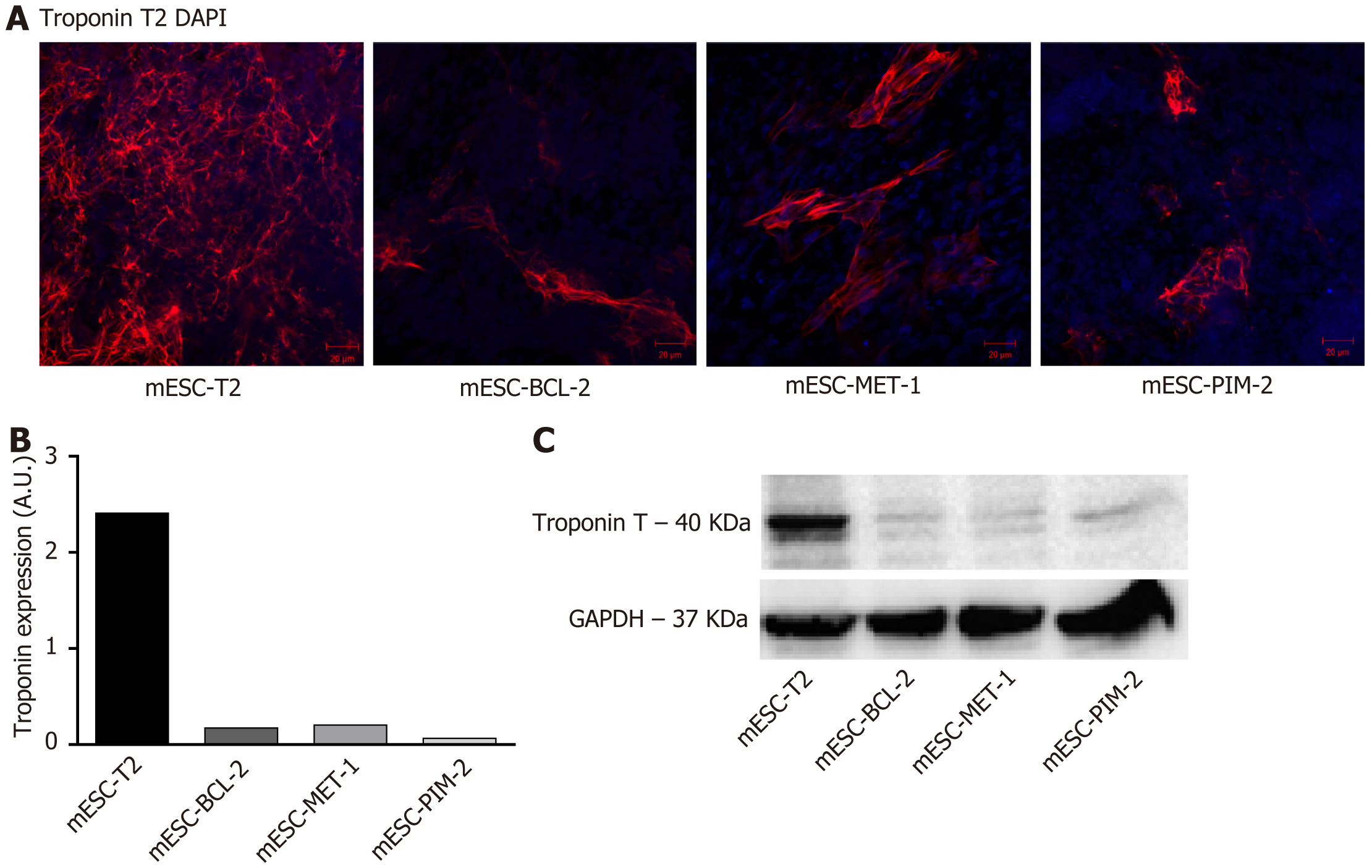
Figure 4 B-cell lymphoma 2, metallothionein-1, and Pim-2 overexpression suppresses the protein expression of troponin T in mouse embryonic stem cells.
Lysates of mouse embryonic stem cells were analyzed using immunofluorescence staining and western blot analysis. A: Representative immunofluorescence images stained with troponin T antibody (red) and the nuclear counterstain antifade Fluorogel II with 4’,6-diamidino-2-phenylindole (blue) (20 × magnification, scale bar = 100 μm). The images were acquired using confocal microscopy; B: Quantification of the red fluorescence intensity for troponin T. The quantification was performed using the Carl Zeiss ZEN 2012 image software; C: Immunoblots for troponin T. Bands were detected by enhanced chemiluminescence using the ChemiDoc MP Imaging System. GAPDH served as an internal control. mESC: Mouse embryonic stem cell; BCL-2: B-cell lymphoma 2; MET-1: Metallothionein-1; DAPI: 4’,6-diamidino-2-phenylindole.
- Citation: Yehya A, Azar J, Al-Fares M, Boeuf H, Abou-Kheir W, Zeineddine D, Hadadeh O. Cardiac differentiation is modulated by anti-apoptotic signals in murine embryonic stem cells. World J Stem Cells 2024; 16(5): 551-559
- URL: https://www.wjgnet.com/1948-0210/full/v16/i5/551.htm
- DOI: https://dx.doi.org/10.4252/wjsc.v16.i5.551


