Copyright
©The Author(s) 2024.
World J Stem Cells. May 26, 2024; 16(5): 538-550
Published online May 26, 2024. doi: 10.4252/wjsc.v16.i5.538
Published online May 26, 2024. doi: 10.4252/wjsc.v16.i5.538
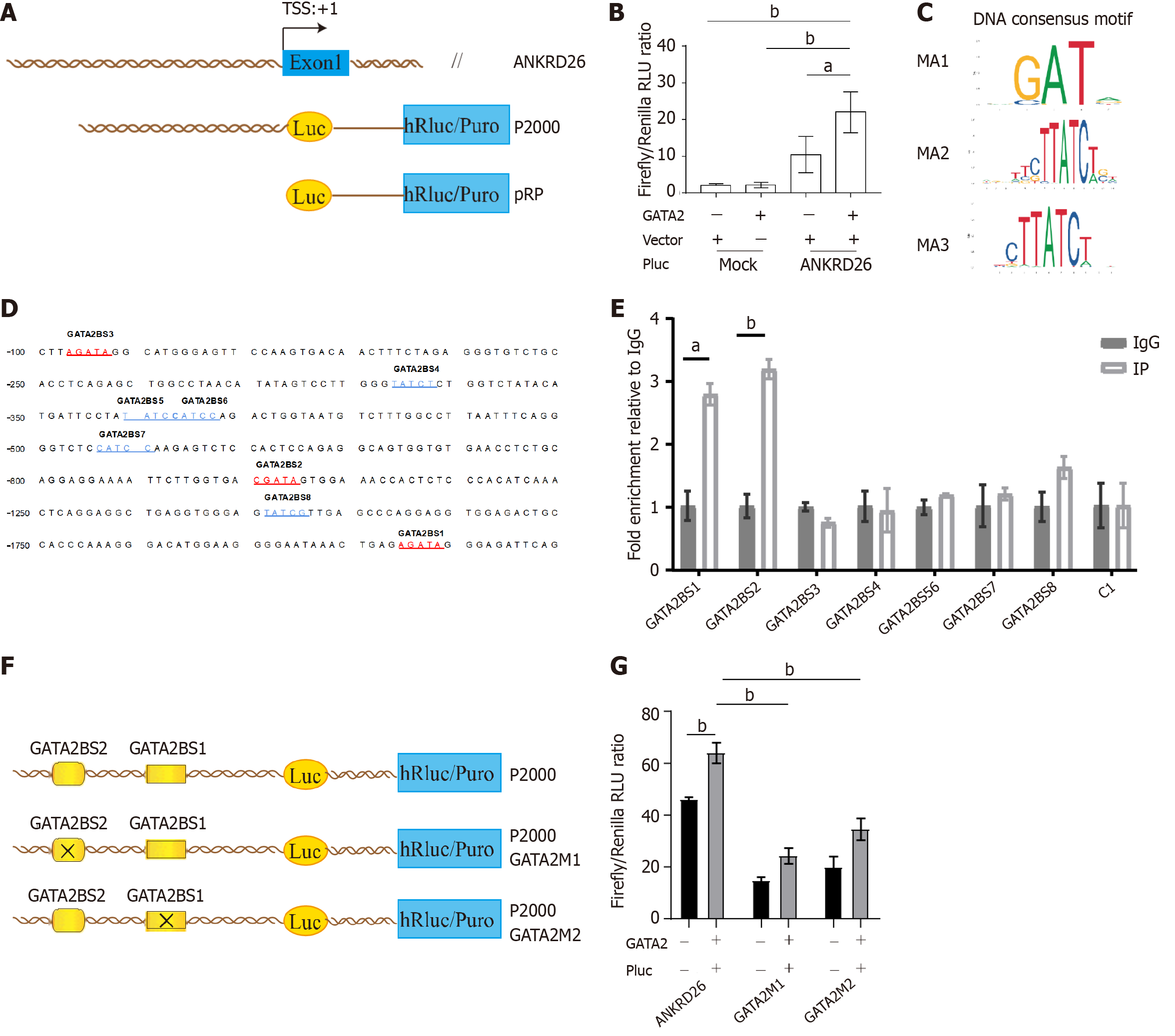
Figure 5 GATA binding protein 2 promotes ankyrin repeat domain containing 26 expression by binding to its promoter region.
A: Schematic diagram of the ankyrin repeat domain containing 26 (ANKRD26) gene promoter reporter constructs. The constructs are named P-length. The open box shows the first exon of ANKRD26, and the positions relative to the major ANKRD26 transcription start site (+1) are indicated. The pRP represents an empty vector (mock); B: The empty (mock) or ANKRD26 (P2000) dual-luciferase reporter constructs were cotransfected into K562 cells with control plasmids or plasmids expressing GATA binding protein 2 (GATA2) in synergy. Cell extracts were analyzed for luciferase activity; C: DNA consensus motifs of GATA2; D: The eight potential GATA2 binding sites on the ANKRD26 promoter region. Red-marked sites are on the sense strand, and blue-marked sites are on the antisense strand; E: Chromatin immunoprecipitation assays performed in K562 cells show that GATA2 directly binds to the ANKRD26 promoter region. The binding sites of GATA2BS1 to GATA2BS8 encompass the 8 predicted binding sites in (D). C1, which encompasses the region without GATA2 binding sites, was used as the negative control; F: Schematic diagram of site-directed mutagenesis of GATA2 binding sites in the ANKRD26 promoter. The two potential GATA2 binding sites are indicated as open boxes (GATA2BS1 and GATA2BS2). The indicated point mutation is denoted by a cross; G: Dual-luciferase assays showing that two mutated GATA2 binding sites block GATA2 Luciferase-promoting activity. The error bars represent the means ± SDs of triplicate samples. aP < 0.05; bP < 0.01, calculated by Student’s t test. GATA2: GATA binding protein 2.
- Citation: Jiang YZ, Hu LY, Chen MS, Wang XJ, Tan CN, Xue PP, Yu T, He XY, Xiang LX, Xiao YN, Li XL, Ran Q, Li ZJ, Chen L. GATA binding protein 2 mediated ankyrin repeat domain containing 26 high expression in myeloid-derived cell lines. World J Stem Cells 2024; 16(5): 538-550
- URL: https://www.wjgnet.com/1948-0210/full/v16/i5/538.htm
- DOI: https://dx.doi.org/10.4252/wjsc.v16.i5.538


