Copyright
©The Author(s) 2024.
World J Stem Cells. May 26, 2024; 16(5): 525-537
Published online May 26, 2024. doi: 10.4252/wjsc.v16.i5.525
Published online May 26, 2024. doi: 10.4252/wjsc.v16.i5.525
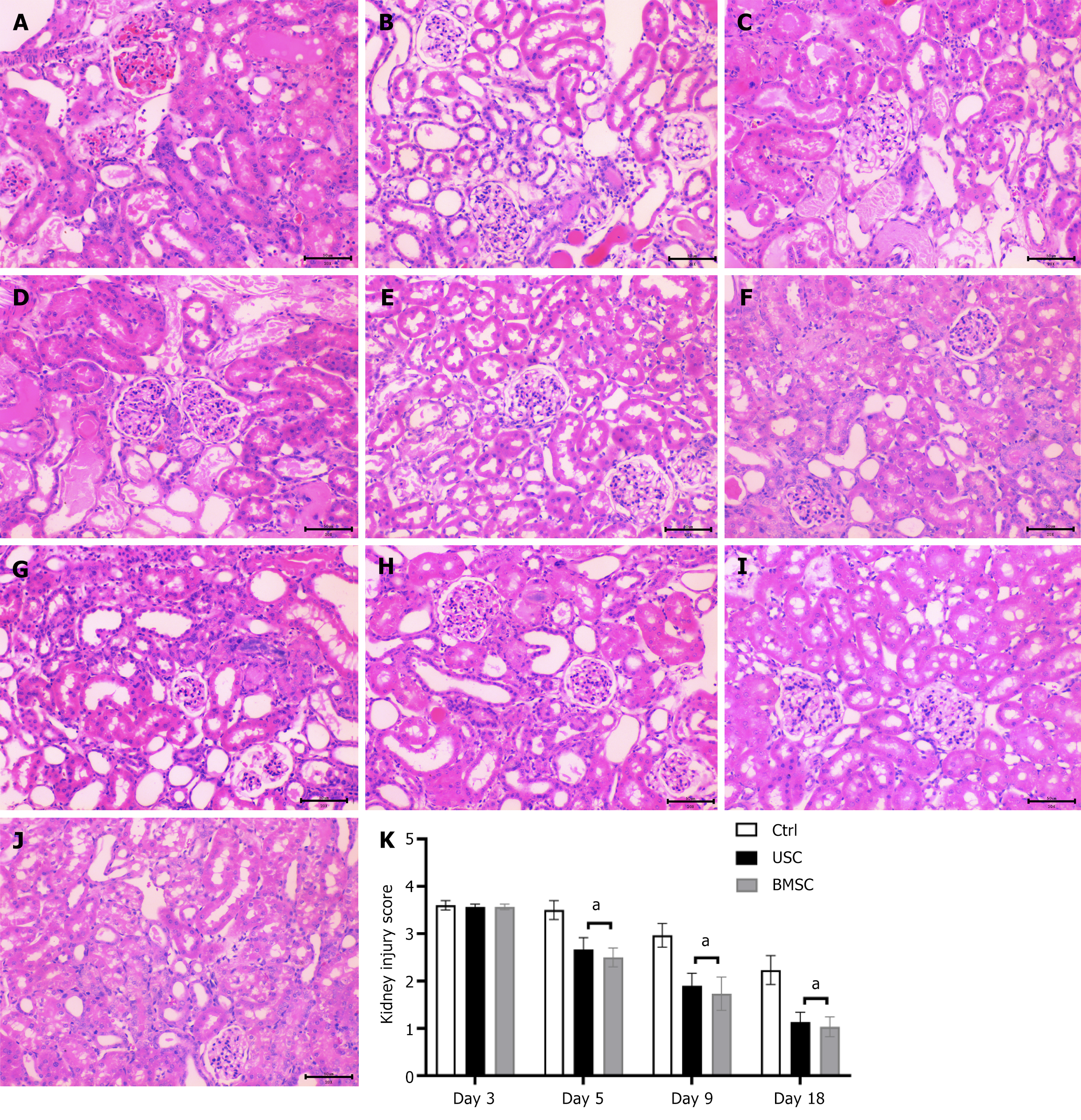
Figure 6 Histological findings of acute kidney injury by hematoxylin and eosin staining (200 ×) in urine-derived stem cell-treated, bone marrow mesenchymal stem cell-treated and control untreated mice.
A-D: Representative micrographs of renal tissue from mice on day 3 (A) and day 5 (D) after the glycerol injection procedure, demonstrating tubular dilatation (red asterisk), tubular necrosis (green arrows), tubular protein cast formation (black asterisk), dilatation of Bowman’s capsule (blue arrows) and loss of the brush border in renal tubules (yellow arrows). Representative micrographs of renal tissue from mice on day 5 post-glycerol injection treated with urine-derived stem cells (USCs) (B) and bone marrow mesenchymal stem cells (BMSCs) (C), showing signs of tissue injury recovery; E-G: Representative micrographs of renal tissue from mice on day 9 post-glycerol injection, exhibiting the persistence of renal injury in untreated mice (G) and signs of recovery in USC-treated (E) or BMSC-treated (F) mice; H-J: Representative micrographs of renal tissue from mice on day 18 showing the normal morphology of tissue in mice untreated (H) or treated with USCs (I) and BMSCs (J). Scale bars in the lower right corner represent 50 μm; K: Graphs illustrating the quantification of kidney injury scores at days 3, 5, 9, and 18 in acute kidney injury mice treated with USCs or BMSCs or injected with saline as a control. Statistical significance was calculated using analysis of variance with the Newman-Keuls multiple comparison test: aP < 0.05, stem cells in acute kidney injury-treated versus untreated mice. USC: Urine-derived stem cell; BMSC: Bone marrow mesenchymal stem cell.
- Citation: Li F, Zhao B, Zhang L, Chen GQ, Zhu L, Feng XL, Gong MJ, Hu CC, Zhang YY, Li M, Liu YQ. Therapeutic potential of urine-derived stem cells in renal regeneration following acute kidney injury: A comparative analysis with mesenchymal stem cells. World J Stem Cells 2024; 16(5): 525-537
- URL: https://www.wjgnet.com/1948-0210/full/v16/i5/525.htm
- DOI: https://dx.doi.org/10.4252/wjsc.v16.i5.525


