Copyright
©The Author(s) 2024.
World J Stem Cells. May 26, 2024; 16(5): 499-511
Published online May 26, 2024. doi: 10.4252/wjsc.v16.i5.499
Published online May 26, 2024. doi: 10.4252/wjsc.v16.i5.499
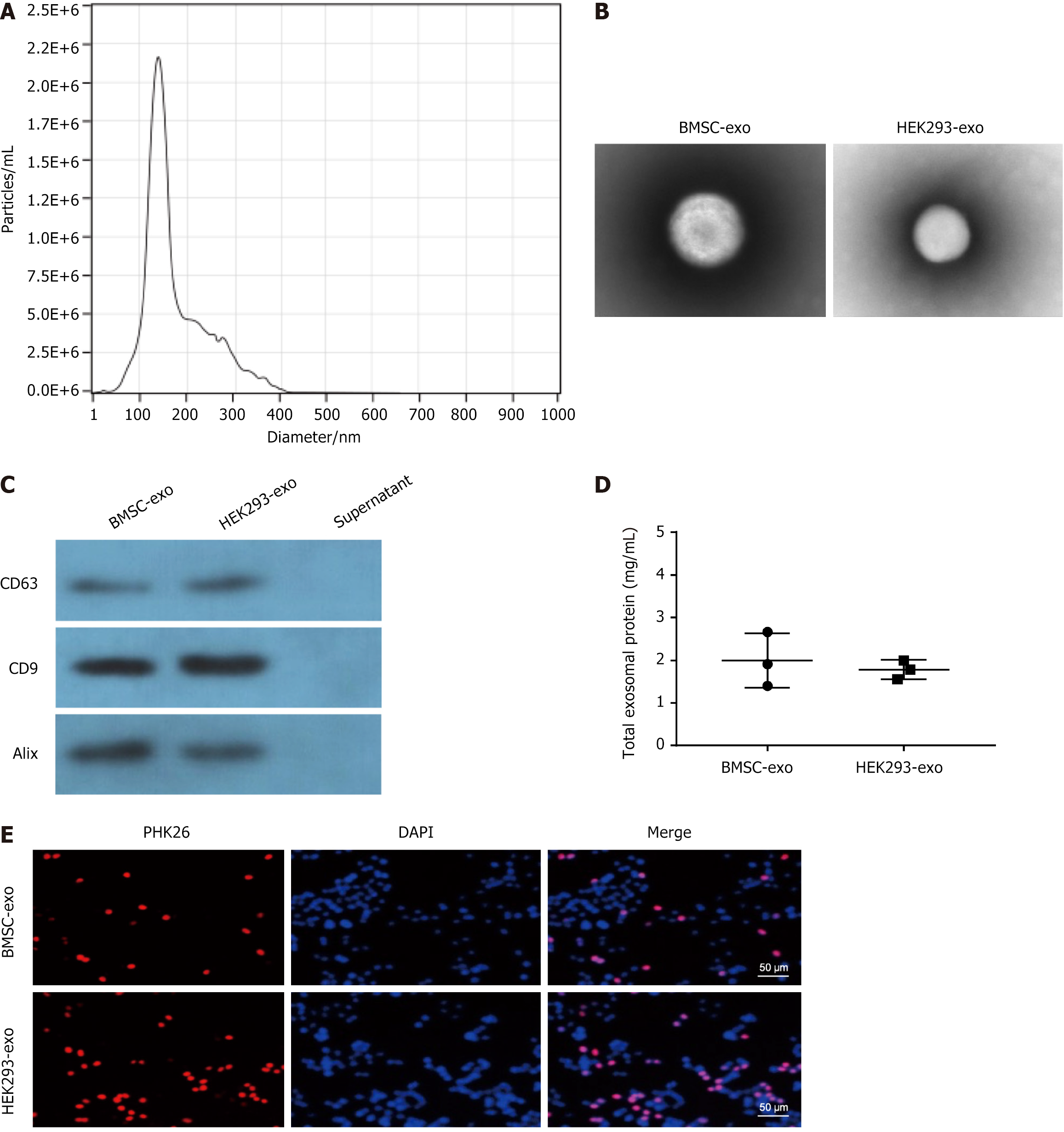
Figure 1 Characterization of bone marrow-derived mesenchymal stem cell- and HEK293-derived exosomes.
A: Particle size distribution of bone marrow-derived mesenchymal stem cell (BMSC)-derived and HEK293-derived exosomes (BMSC-exo and HEK293-exo, respectively) detected by NanoSight; the mean diameter is 150 nm; B: Representative images of the morphology of BMSC-exo and HEK293-exo via transmission electron microscopy; C: Western blot identification of exosome surface markers; D: Expression of BMSC-exo and HEK293-exo protein; E: Internalization of PKH26-labeled exosomes within mouse osteoblast progenitor cells by fluorescence microscopy, Scale bar: 50 μm. BMSC-exo: Bone marrow-derived mesenchymal stem cell-derived exosome.
- Citation: Zhang S, Lu C, Zheng S, Hong G. Hydrogel loaded with bone marrow stromal cell-derived exosomes promotes bone regeneration by inhibiting inflammatory responses and angiogenesis. World J Stem Cells 2024; 16(5): 499-511
- URL: https://www.wjgnet.com/1948-0210/full/v16/i5/499.htm
- DOI: https://dx.doi.org/10.4252/wjsc.v16.i5.499


