Copyright
©The Author(s) 2024.
World J Stem Cells. Apr 26, 2024; 16(4): 434-443
Published online Apr 26, 2024. doi: 10.4252/wjsc.v16.i4.434
Published online Apr 26, 2024. doi: 10.4252/wjsc.v16.i4.434
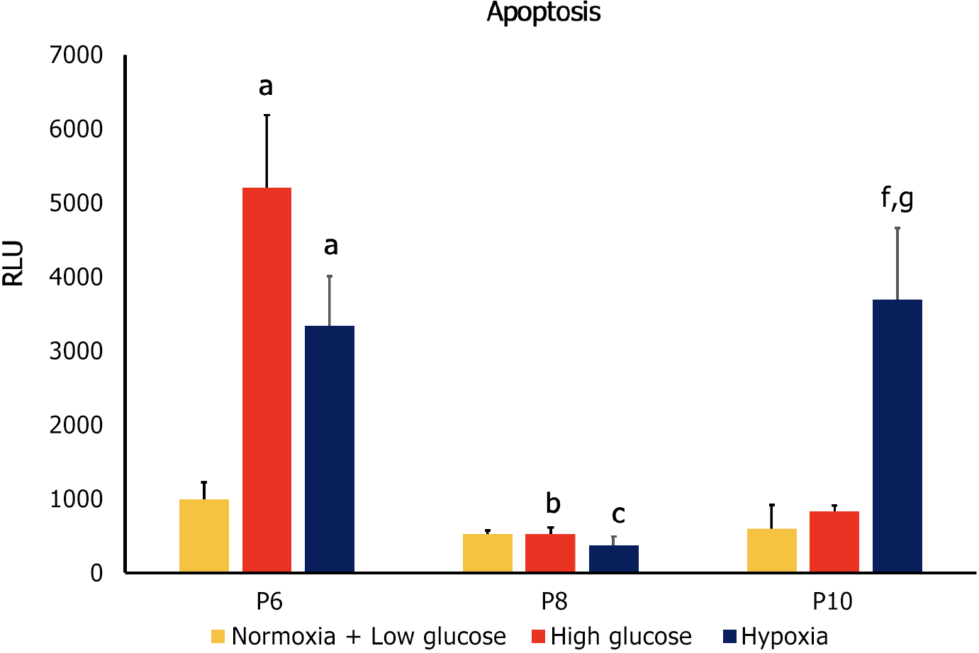
Figure 5 Apoptosis of human adipose-tissue derived mesenchymal stem cells at passages 6, 8 and 10 under conditions of normoxia + low glucose (control), high glucose and hypoxia, as indicated by Annexin V.
Mesenchymal stem cells (MSCs) at passages 6 (P6) showed increased apoptosis under conditions of high glucose and hypoxia compared to control cells. At P8 apoptosis significantly decreased in both stress conditions compared to their counterparts at P6, while no significant difference was detected compared to the control group at the same passage. At P10 only cells cultured in hypoxia had a significantly higher apoptosis compared to control group at P10 and to counterpart cells at both P6 and P8. aP < 0.05 vs passages 6 (P6) normoxia + low glucose; bP < 0.05 vs P6 high glucose; cp < 0.05 vs P6 hypoxia; fP < 0.05 compared to P8 hypoxia; gP < 0.05 compared to P10 normoxia + low glucose. P6: Passage 6; P8: Passage 8; P10: Passage 10; RLU: Relative luminescence intensity.
- Citation: Almahasneh F, Abu-El-Rub E, Khasawneh RR, Almazari R. Effects of high glucose and severe hypoxia on the biological behavior of mesenchymal stem cells at various passages. World J Stem Cells 2024; 16(4): 434-443
- URL: https://www.wjgnet.com/1948-0210/full/v16/i4/434.htm
- DOI: https://dx.doi.org/10.4252/wjsc.v16.i4.434


