Copyright
©The Author(s) 2024.
World J Stem Cells. Apr 26, 2024; 16(4): 389-409
Published online Apr 26, 2024. doi: 10.4252/wjsc.v16.i4.389
Published online Apr 26, 2024. doi: 10.4252/wjsc.v16.i4.389
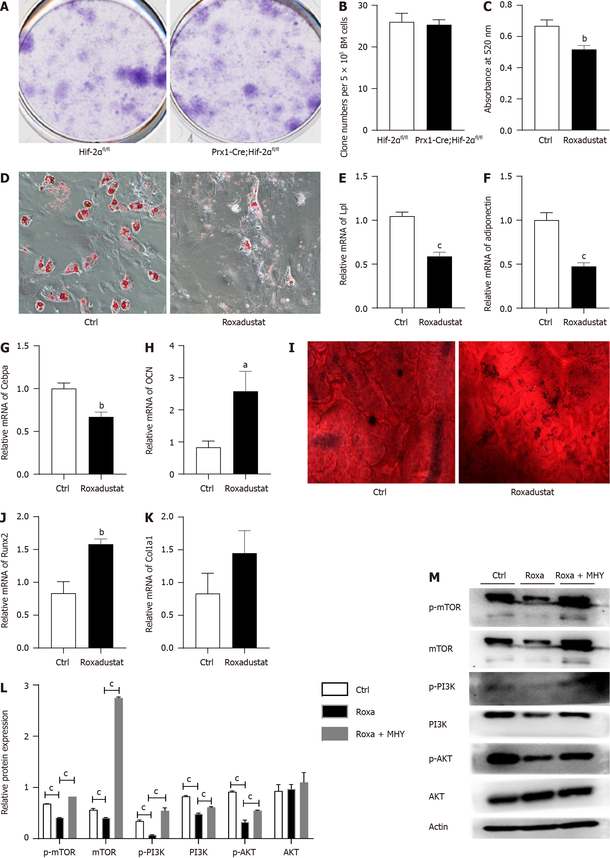
Figure 6 Treatment with the hypoxia-inducible factor 2alpha agonist roxadustat increases osteogenesis and decreases adipogenesis.
A: Representative images of the fibroblast colony-forming unit (CFU-F) assay with Prx1-Cre;Hif-2αfl/fl and Hif-2αfl/fl mice are shown; B: Quantification of CFU-Fs formed by bone mesenchymal stem cells (BMSCs) from Prx1-Cre;Hif-2αfl/fl and Hif-2αfl/fl mice are shown; C and D: Hif-2αfl/fl mouse BMSCs were cultured with or without roxadustat. In the adipogenic differentiation assay, Oil red O staining was performed after seven days (D); quantification of Oil Red O staining in BMSCs after induction of adipogenic differentiation (C); E-M: Relative mRNA levels of the adipogenesis-related genes Lpl (E), Adiponectin (F) and Cebpa (G) in BMSCs after 7 d of adipogenic differentiation. In the osteogenic differentiation assay, Alizarin red staining was performed after 14 d to quantify osteoblast mineralization (I). Relative mRNA levels of the osteogenesis-related genes OCN (H), Runx2 (J) and Col1α1 (K) in BMSCs after 14 d of osteogenic differentiation. Adipogenic differentiation was induced in BMSCs by treatment with roxadustat and MHY1485 for 7 d. The levels of the mechanistic target of rapamycin (mTOR) and PI3K/AKT signaling pathway proteins were measured via western blotting in the Ctrl, roxadustat and Roxadustat + MHY1485 groups (M). Quantification of the relative levels of the mTOR, PI3K and AKT proteins (L). The phosphorylated protein levels were normalized to the corresponding total protein levels. aP < 0.05, bP < 0.01, cP < 0.001. Roxa: Roxadustat; MHY: MHY1485; HIF-2α: Hypoxia-inducible factor 2alpha; mTOR: Mechanistic target of rapamycin.
- Citation: Wang LL, Lu ZJ, Luo SK, Li Y, Yang Z, Lu HY. Unveiling the role of hypoxia-inducible factor 2alpha in osteoporosis: Implications for bone health. World J Stem Cells 2024; 16(4): 389-409
- URL: https://www.wjgnet.com/1948-0210/full/v16/i4/389.htm
- DOI: https://dx.doi.org/10.4252/wjsc.v16.i4.389


