Copyright
©The Author(s) 2024.
World J Stem Cells. Apr 26, 2024; 16(4): 389-409
Published online Apr 26, 2024. doi: 10.4252/wjsc.v16.i4.389
Published online Apr 26, 2024. doi: 10.4252/wjsc.v16.i4.389
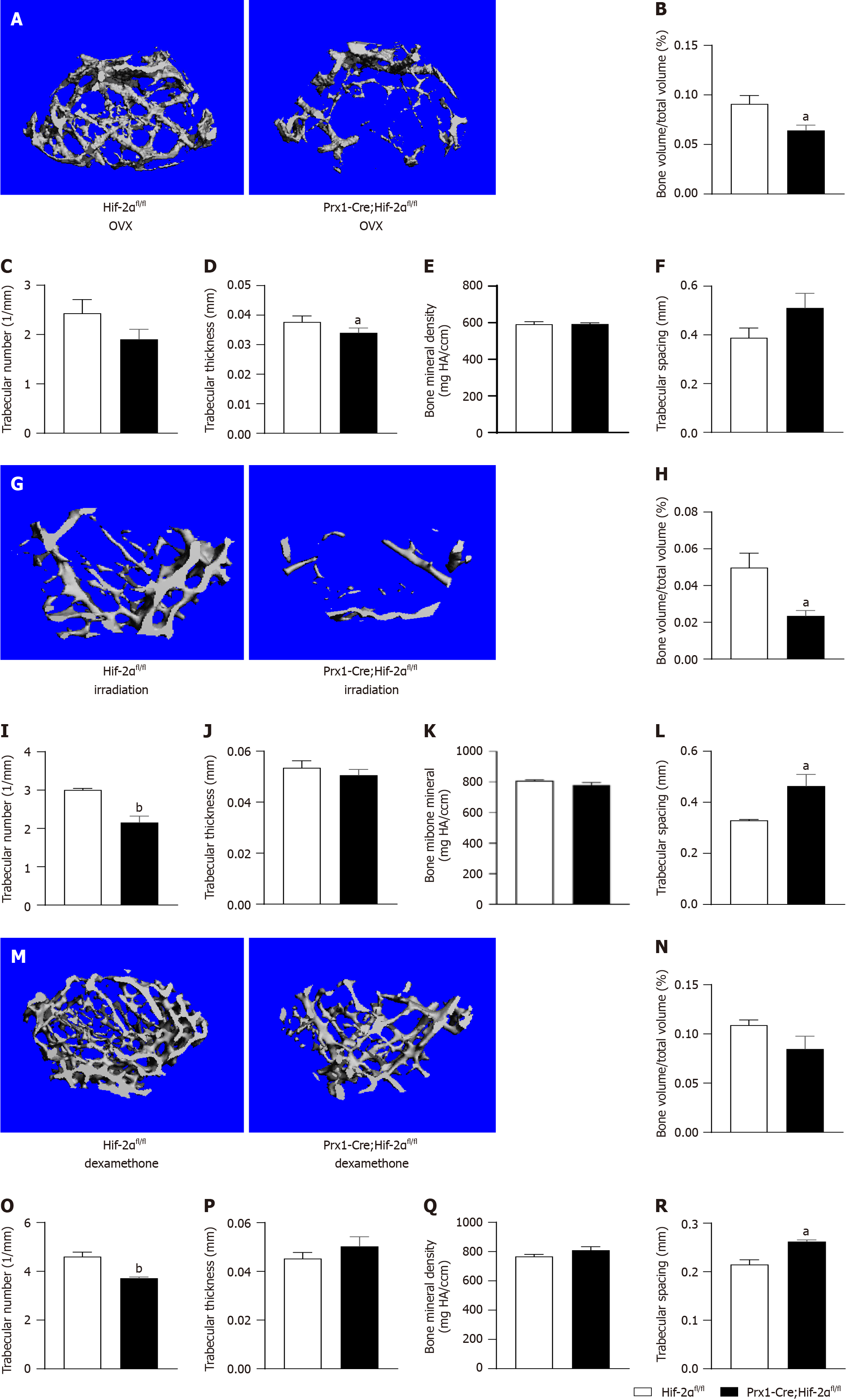
Figure 4 Adult Prx1-Cre;Hif-2αfl/fl and Hif-2αfl/fl mice exhibit decreased bone mass under three different stimulation conditions.
A: Representative microcomputed tomography (μCT) images of femora from both Prx1-Cre;Hif-2αfl/fl and Hif-2αfl/fl mice 8 wk after ovariectomy; B-E: The bone volume (BV) fraction [BV/total volume (TV)] (B), trabecular number (Tb.N) (C), trabecular thickness (Tb.Th) (D) and bone mineral density (E) in the distal metaphysis of the femur were decreased by ovariectomy in Prx1-Cre;Hif-2αfl/fl mice, while the trabecular spacing (Tb.Sp) was appreciably increased in Prx1-Cre;Hif-2αfl/fl mice; G: Representative μCT images of femora from both Prx1-Cre;Hif-2αfl/fl and Hif-2αfl/fl mice 4 wk after semilethal irradiation; H-L: The BV/TV (H), Tb.N (I), Tb.Th (J) and BMD (K) in the distal metaphysis of the femur were obviously decreased by semilethal irradiation in Prx1-Cre;Hif-2αfl/fl mice, while the Tb.Sp (L) was appreciably increased in Prx1-Cre;Hif-2αfl/fl mice; M: Representative μCT images of femora from both Prx1-Cre;Hif-2αfl/fl and Hif-2αfl/fl mice 4 wk after dexamethasone treatment; N-R: The BV/TV (N), Tb.N (O), Tb.Th (P) and BMD (Q) in the distal metaphysis of the femur were obviously decreased by dexamethasone treatment in Prx1-Cre;Hif-2αfl/fl mice, while the Tb.Sp (R) was appreciably increased in Prx1-Cre;Hif-2αfl/fl mice. aP < 0.05, bP < 0.01.
- Citation: Wang LL, Lu ZJ, Luo SK, Li Y, Yang Z, Lu HY. Unveiling the role of hypoxia-inducible factor 2alpha in osteoporosis: Implications for bone health. World J Stem Cells 2024; 16(4): 389-409
- URL: https://www.wjgnet.com/1948-0210/full/v16/i4/389.htm
- DOI: https://dx.doi.org/10.4252/wjsc.v16.i4.389


