Copyright
©The Author(s) 2024.
World J Stem Cells. Apr 26, 2024; 16(4): 389-409
Published online Apr 26, 2024. doi: 10.4252/wjsc.v16.i4.389
Published online Apr 26, 2024. doi: 10.4252/wjsc.v16.i4.389
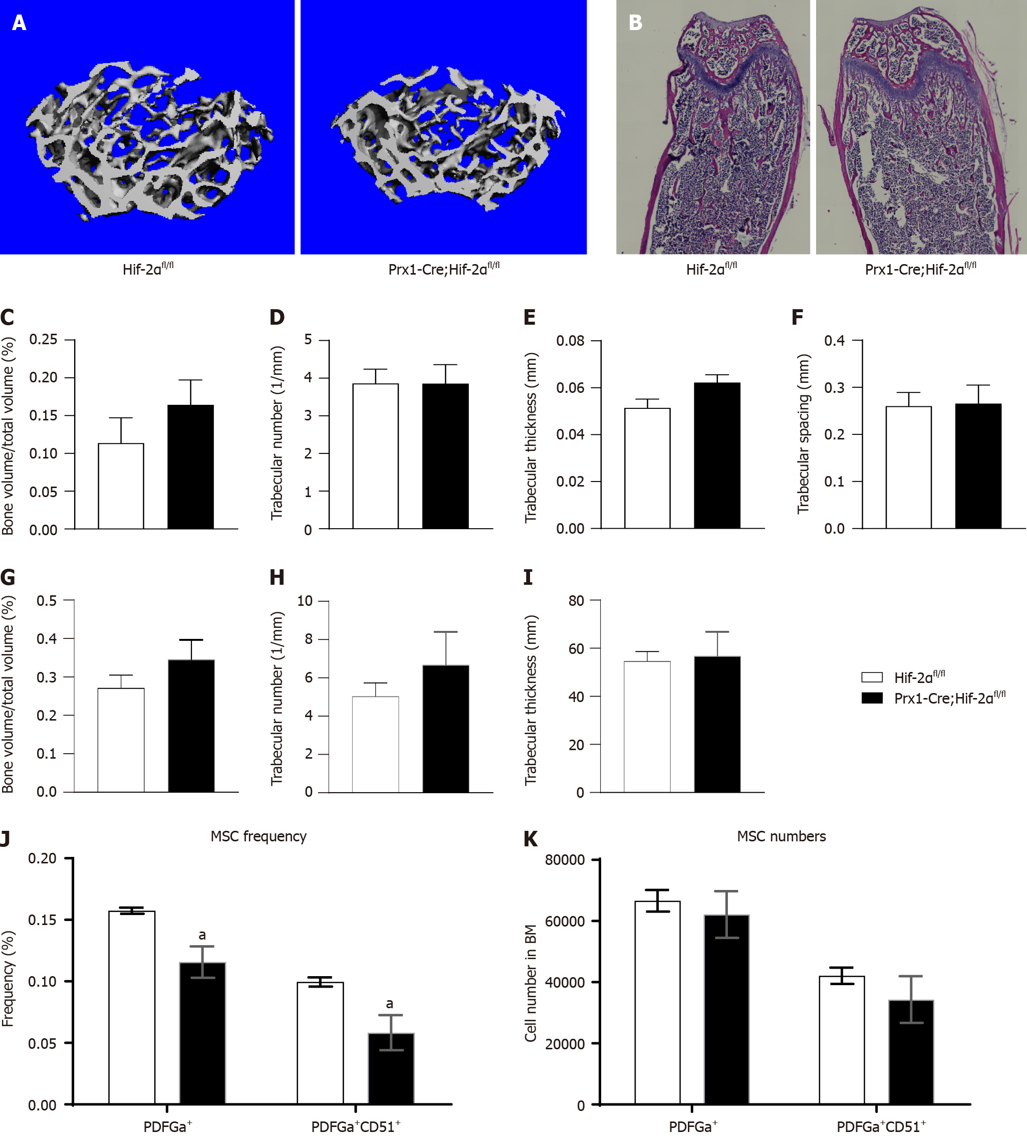
Figure 3 Adult Prx1-Cre;Hif-2αfl/fl and Hif-2αfl/fl mice exhibit no difference in bone mass under the naive condition.
A: Microcomputed tomography (μCT) reconstruction images of distal femoral metaphyses from 2-month-old Prx1-Cre;Hif-2αfl/fl and Hif-2αfl/fl mice; B: Hematoxylin and eosin (HE) staining images of distal femoral metaphyses from 2-month-old Prx1-Cre;Hif-2αfl/fl and Hif-2αfl/fl mice; C-K: Quantitative μCT analyses of the structural parameters of femoral trabeculae from 2-month-old mice: bone volume/tissue volume (C), trabecular number (D), trabecular thickness (E), and trabecular spacing (F); quantitative HE staining analysis of the structural parameters of femoral trabeculae from 2-month-old mice: bone volume/tissue volume (G), trabecular number (H), and trabecular thickness (I); bone marrow cells freshly harvested from 8-wk-old Prx1-Cre;Hif-2αfl/fl and Hif-2αfl/fl mice were assayed by multiparameter fluorescence-activated cell sorting analyses to determine the frequency (J) and number (K) of bone mesenchymal stem cells. aP < 0.05. MSC: Mesenchymal stem cell.
- Citation: Wang LL, Lu ZJ, Luo SK, Li Y, Yang Z, Lu HY. Unveiling the role of hypoxia-inducible factor 2alpha in osteoporosis: Implications for bone health. World J Stem Cells 2024; 16(4): 389-409
- URL: https://www.wjgnet.com/1948-0210/full/v16/i4/389.htm
- DOI: https://dx.doi.org/10.4252/wjsc.v16.i4.389


