Copyright
©The Author(s) 2024.
World J Stem Cells. Mar 26, 2024; 16(3): 287-304
Published online Mar 26, 2024. doi: 10.4252/wjsc.v16.i3.287
Published online Mar 26, 2024. doi: 10.4252/wjsc.v16.i3.287
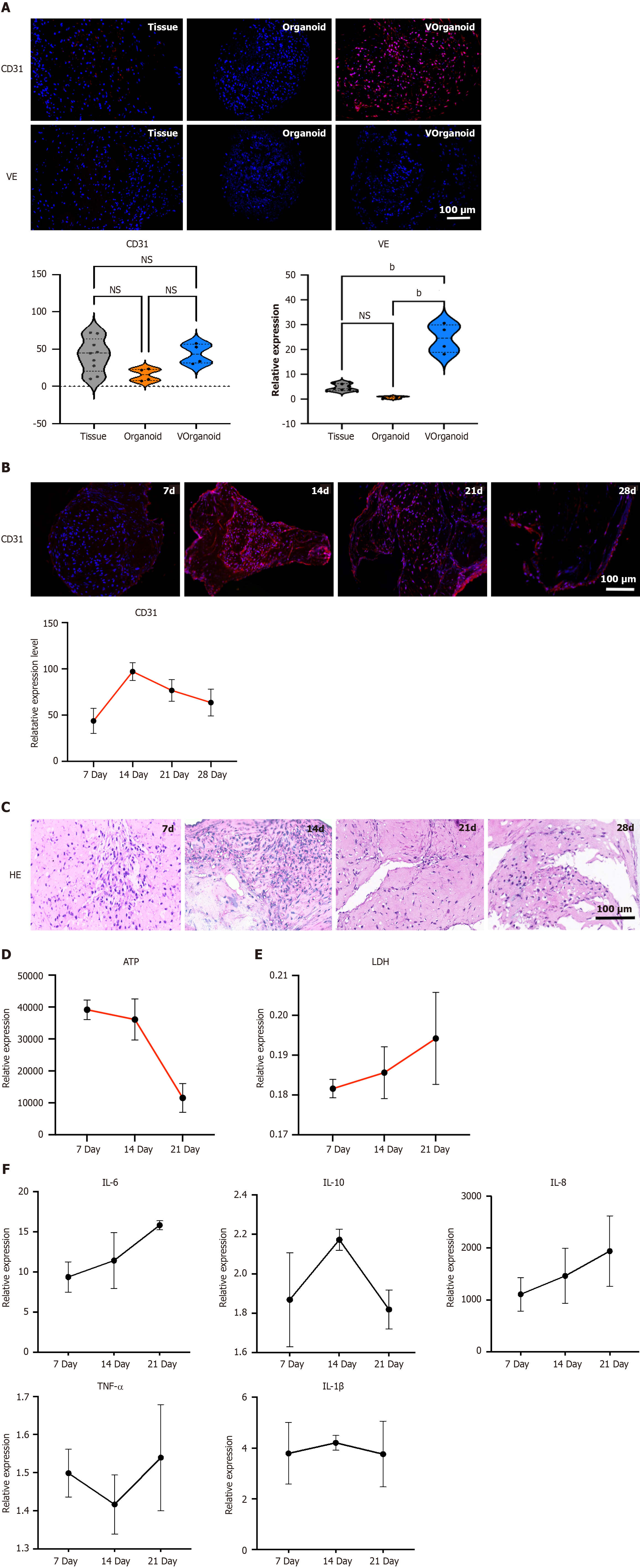
Figure 4 Morphological and functional identification of Vorganoids.
A: Immunofluorescence staining for vasculogenesis-related markers; B: CD31 expression pattern in Vorganoid cells during different culture durations; C: Morphological changes in Vorganoid cells during different culture durations; D and E: Analysis of adenosine triphosphate (D) and lactate dehydrogenase (E) levels in Vorganoids during different culture durations; F: Expression patterns of inflammatory markers in Vorganoids analysed via enzyme-linked immunosorbent assay. Significant difference between the groups, bP < 0.01, Scale bar = 100 μm. HE: Hematoxylin-eosin; ATP: Adenosine triphosphate; LDH: Lactate dehydrogenase; IL: Interleukin; TNF: Tumour necrosis factor; NS: Not significant.
- Citation: Liu F, Xiao J, Chen LH, Pan YY, Tian JZ, Zhang ZR, Bai XC. Self-assembly of differentiated dental pulp stem cells facilitates spheroid human dental organoid formation and prevascularization. World J Stem Cells 2024; 16(3): 287-304
- URL: https://www.wjgnet.com/1948-0210/full/v16/i3/287.htm
- DOI: https://dx.doi.org/10.4252/wjsc.v16.i3.287


