Copyright
©The Author(s) 2024.
World J Stem Cells. Mar 26, 2024; 16(3): 267-286
Published online Mar 26, 2024. doi: 10.4252/wjsc.v16.i3.267
Published online Mar 26, 2024. doi: 10.4252/wjsc.v16.i3.267
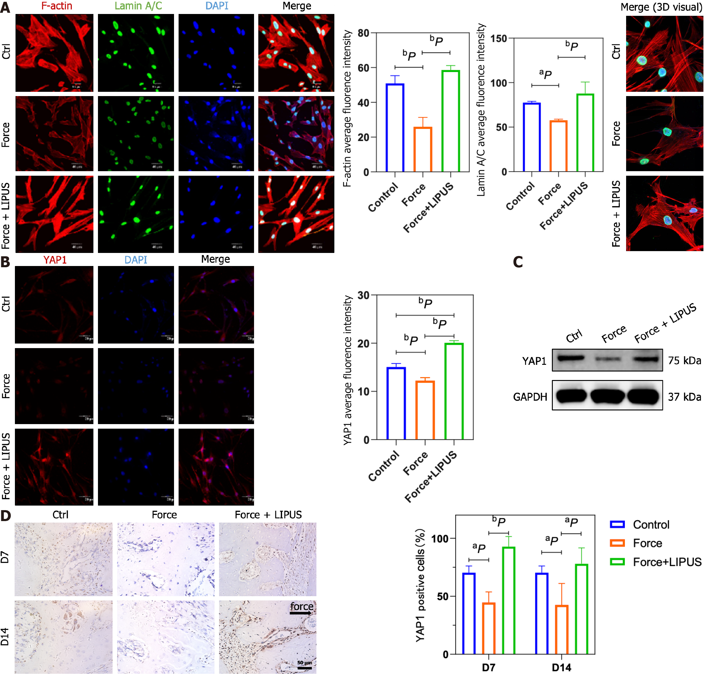
Figure 6 Low-intensity pulsed ultrasound reverses the stress-induced decrease in LaminA/C, F-actin, and Yes-associated protein expression.
A: Representative immunofluorescence images of F-actin (red), LaminA/C (green), and DAPI (blue) staining (left) and statistical analyses (middle), as well as representative 3D merged images constructed with Imaris software (right); B: Representative immunofluorescence images of Yes-associated protein (YAP1) (red) and DAPI (blue) staining (left) and statistical analyses (right); C: YAP1 protein expression analyzed by Western blotting; D: Representative images of immunohistochemical staining for YAP1 (left) and statistical analysis (right). aP < 0.05 vs control group, bP < 0.01 vs control group. LIPUS: Low-intensity pulsed ultrasound; YAP1: Yes-associated protein.
- Citation: Wu T, Zheng F, Tang HY, Li HZ, Cui XY, Ding S, Liu D, Li CY, Jiang JH, Yang RL. Low-intensity pulsed ultrasound reduces alveolar bone resorption during orthodontic treatment via Lamin A/C-Yes-associated protein axis in stem cells. World J Stem Cells 2024; 16(3): 267-286
- URL: https://www.wjgnet.com/1948-0210/full/v16/i3/267.htm
- DOI: https://dx.doi.org/10.4252/wjsc.v16.i3.267


