Copyright
©The Author(s) 2024.
World J Stem Cells. Mar 26, 2024; 16(3): 267-286
Published online Mar 26, 2024. doi: 10.4252/wjsc.v16.i3.267
Published online Mar 26, 2024. doi: 10.4252/wjsc.v16.i3.267
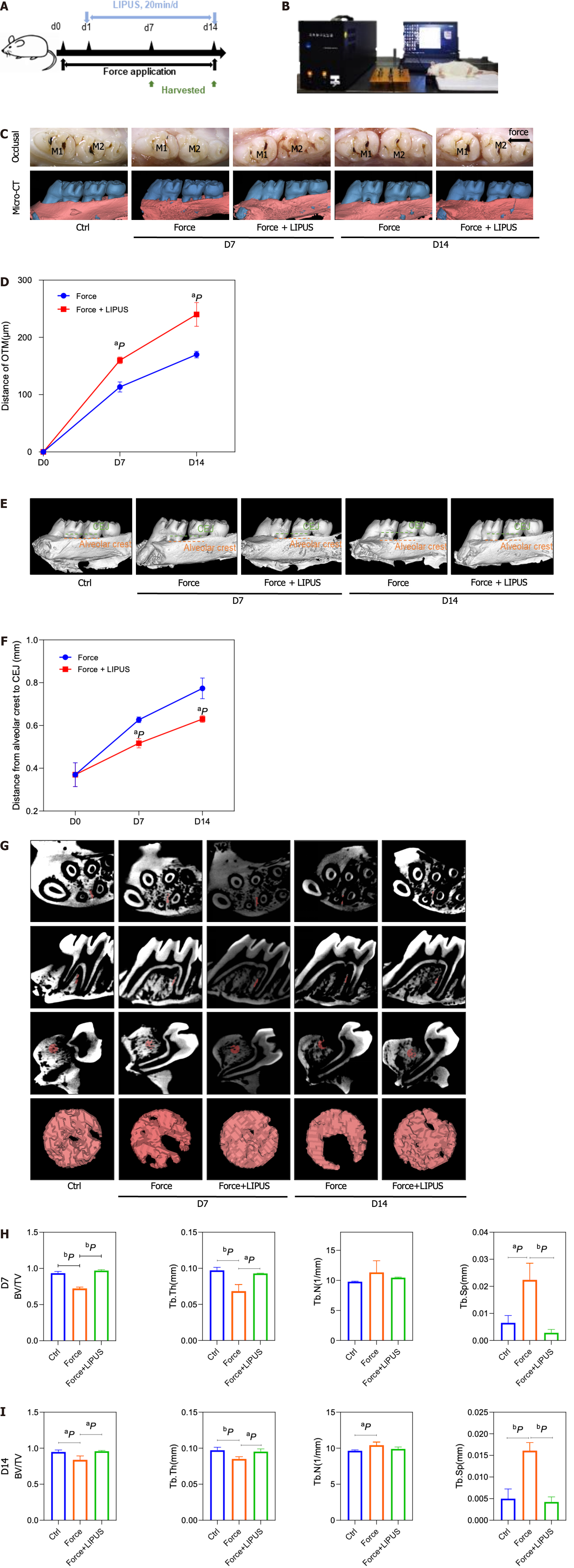
Figure 4 Low-intensity pulsed ultrasound accelerates tooth movement, inhibits alveolar bone resorption, and promotes bone formation.
A: Schematic diagram of the animal experiment procedure. The orthodontic tooth movement model was established on day 0. From day 1 to the end point, the force + low-intensity pulsed ultrasound (LIPUS) group was stimulated with LIPUS for 20 min every day until the samples were harvested on days 7 and 14; B: Schematic representation of LIPUS stimulating the maxillary first molar at the corresponding position on the buccal side; C-F: Representative images (C) and 3D reconstructed model images (E). Tooth movement distances (D) and distances from the alveolar crest to the cementum-enamel boundary (F) were measured with Mimics software; G-I: Representative region of interest (ROI) region selection diagrams and statistical analysis of the BV/TV, Tb.Th, Tb.N, Tb.Sp, and BS/BV values of the ROI. aP < 0.05 vs control group, bP < 0.01 vs control group. CT: Computed tomography; LIPUS: Low-intensity pulsed ultrasound.
- Citation: Wu T, Zheng F, Tang HY, Li HZ, Cui XY, Ding S, Liu D, Li CY, Jiang JH, Yang RL. Low-intensity pulsed ultrasound reduces alveolar bone resorption during orthodontic treatment via Lamin A/C-Yes-associated protein axis in stem cells. World J Stem Cells 2024; 16(3): 267-286
- URL: https://www.wjgnet.com/1948-0210/full/v16/i3/267.htm
- DOI: https://dx.doi.org/10.4252/wjsc.v16.i3.267


