Copyright
©The Author(s) 2024.
World J Stem Cells. Feb 26, 2024; 16(2): 176-190
Published online Feb 26, 2024. doi: 10.4252/wjsc.v16.i2.176
Published online Feb 26, 2024. doi: 10.4252/wjsc.v16.i2.176
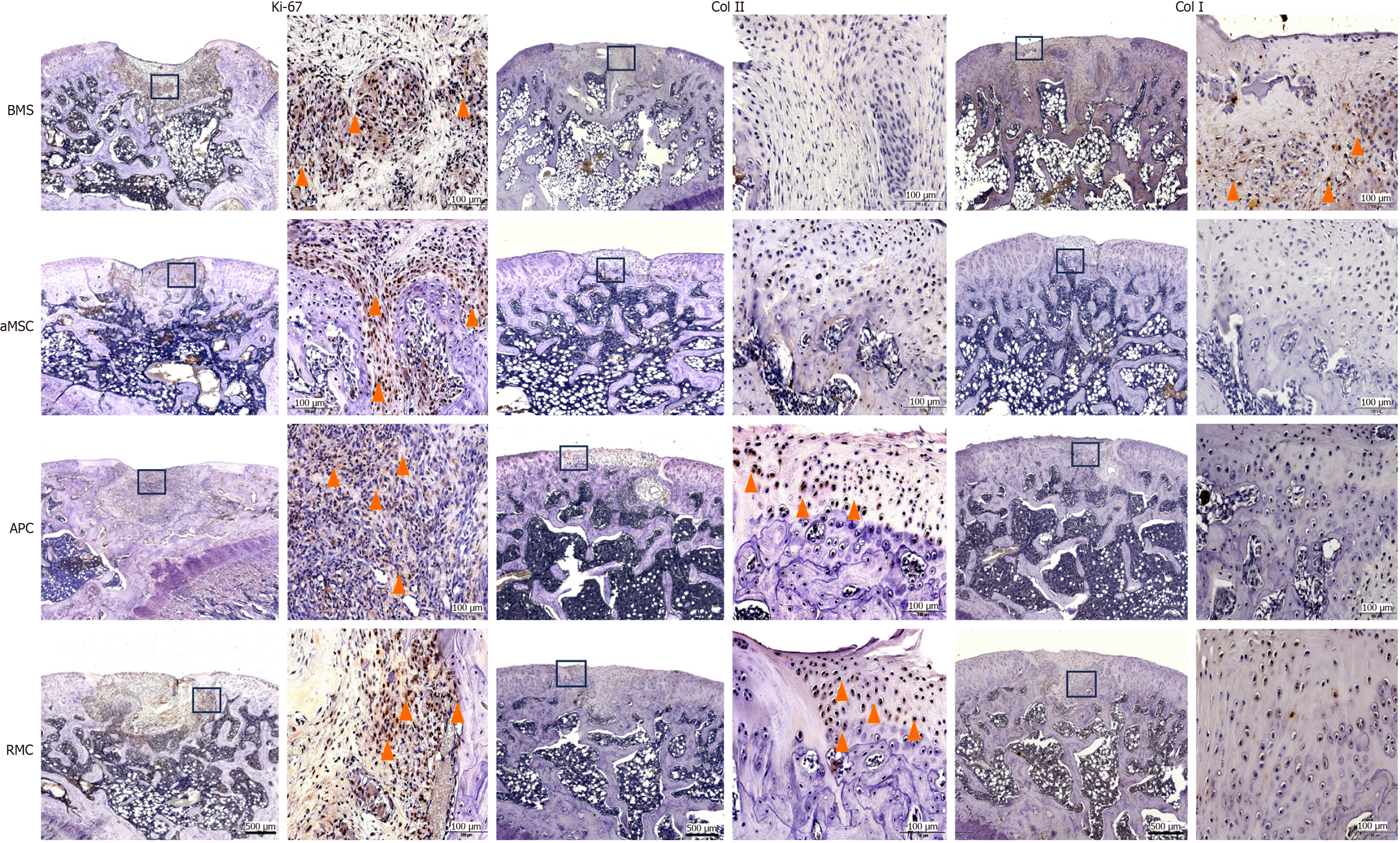
Figure 5 Immunohistochemical evaluation of the top layer covering the osteochondral defects by applying different extracellular matrix-sheets.
Ki-67 staining at week 4: Note that the cells of the re-forming tissue in the defects of all groups were extensively and intensively stained with Ki-67 (yellow arrowheads), indicating that each defect was under active repair. Col II staining at week 12: Note that the surface layer of each defect was positively stained in the antlerogenic periosteal cell (APC) and antler reserve mesenchymal cell (RMC) groups only (yellow arrowheads), but not in the adipocyte-derived mesenchymal stromal cell (aMSC) and bone marrow stimulation (BMS) groups, indicating that the surface tissue in the APC and RMC groups was of cartilage in nature. Col I staining at week 12: Note that the regenerated tissue in the BMS group was significantly positively stained (yellow arrowheads), but that in the other three groups was not, indicating that the surface tissue in the BMS group was more fibrous tissue-like in nature. Bar = 500 μm. BMS: Bone marrow stimulation; aMSC: Adipocyte-derived mesenchymal stromal cell; APC: Antlerogenic periosteal cell; RMC: Antler reserve mesenchymal cell.
- Citation: Wang YS, Chu WH, Zhai JJ, Wang WY, He ZM, Zhao QM, Li CY. High quality repair of osteochondral defects in rats using the extracellular matrix of antler stem cells. World J Stem Cells 2024; 16(2): 176-190
- URL: https://www.wjgnet.com/1948-0210/full/v16/i2/176.htm
- DOI: https://dx.doi.org/10.4252/wjsc.v16.i2.176


