Copyright
©The Author(s) 2024.
World J Stem Cells. Feb 26, 2024; 16(2): 176-190
Published online Feb 26, 2024. doi: 10.4252/wjsc.v16.i2.176
Published online Feb 26, 2024. doi: 10.4252/wjsc.v16.i2.176
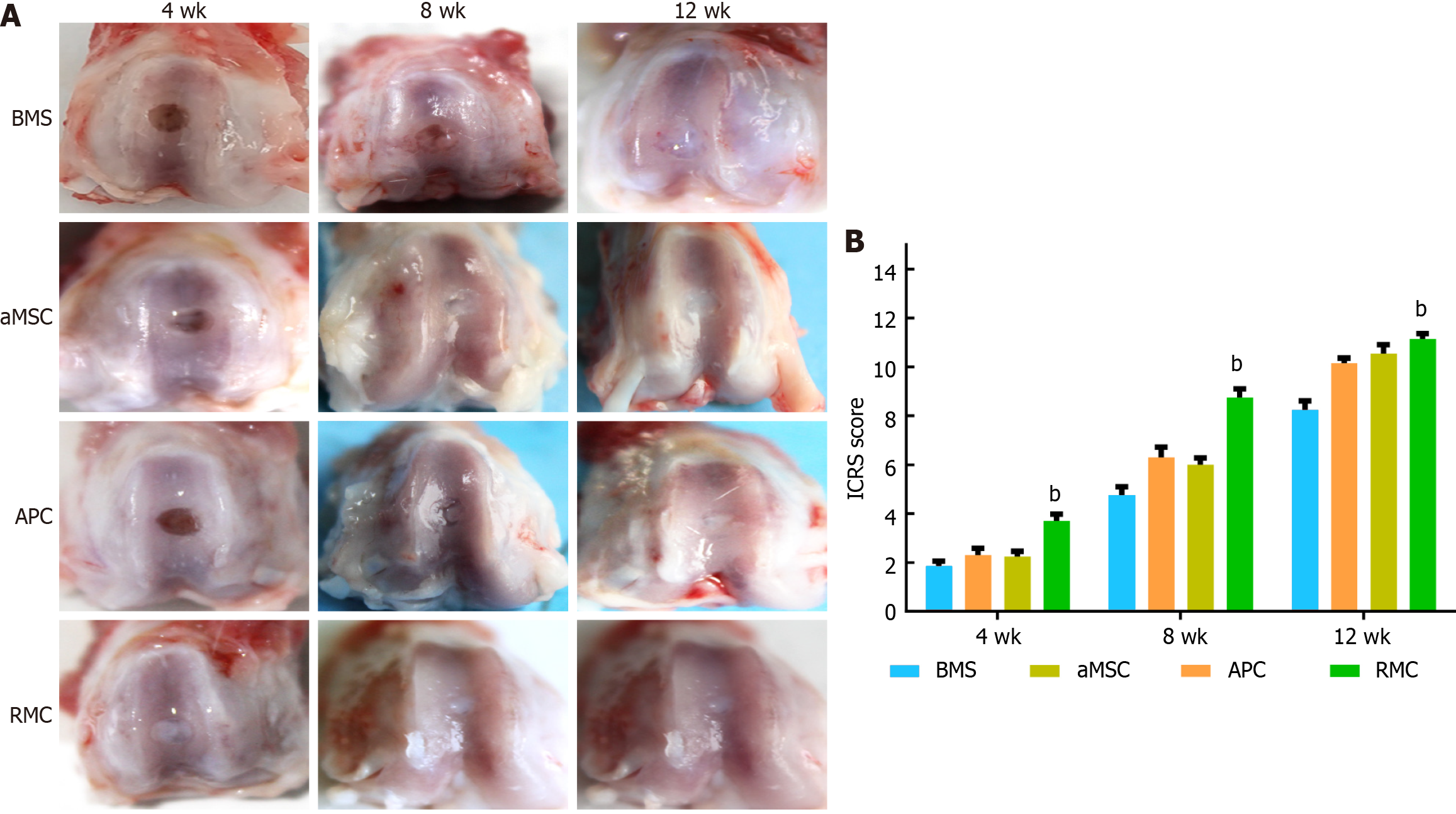
Figure 3 Morphological evaluation and International Cartilage Repair Society scores of the osteochondral defect repair by applying different adipocyte-derived mesenchymal stromal cell-, antlerogenic periosteal cell-, and antler reserve mesenchymal cell-extracellular matrix sheets at 4, 8 and 12 wk.
A: Morphological evaluation; B: International Cartilage Repair Society scores. Note that at week 4, the defect in the antler reserve mesenchymal cell (RMC) group was fully filled with ivory white colored tissue; and in the antlerogenic periosteal cell (APC) and adipocyte-derived mesenchymal stromal cell (aMSC) groups only partially filled with reddish colored tissue; in the bone marrow stimulation (BMS) group, the defect seemed empty and no regenerated tissue was visible. At week 8, the sizes of all the defects in the four groups were substantially reduced, and in the aMSC, RMC and APC groups, they were filled with white colored tissue, although the reduced defect in the BMS group was still partially filled with fibrous-like tissue. At week 12, the defect sizes of all groups were further reduced but to varying degrees, with the RMC group the smallest and BMS and aMSC groups the largest. The International Cartilage Repair Society sores were consistent with the results of morphological observation. bP < 0.01. BMS: Bone marrow stimulation; aMSC: Adipocyte-derived mesenchymal stromal cell; APC: Antlerogenic periosteal cell; RMC: Antler reserve mesenchymal cell.
- Citation: Wang YS, Chu WH, Zhai JJ, Wang WY, He ZM, Zhao QM, Li CY. High quality repair of osteochondral defects in rats using the extracellular matrix of antler stem cells. World J Stem Cells 2024; 16(2): 176-190
- URL: https://www.wjgnet.com/1948-0210/full/v16/i2/176.htm
- DOI: https://dx.doi.org/10.4252/wjsc.v16.i2.176


