Copyright
©The Author(s) 2024.
World J Stem Cells. Feb 26, 2024; 16(2): 176-190
Published online Feb 26, 2024. doi: 10.4252/wjsc.v16.i2.176
Published online Feb 26, 2024. doi: 10.4252/wjsc.v16.i2.176
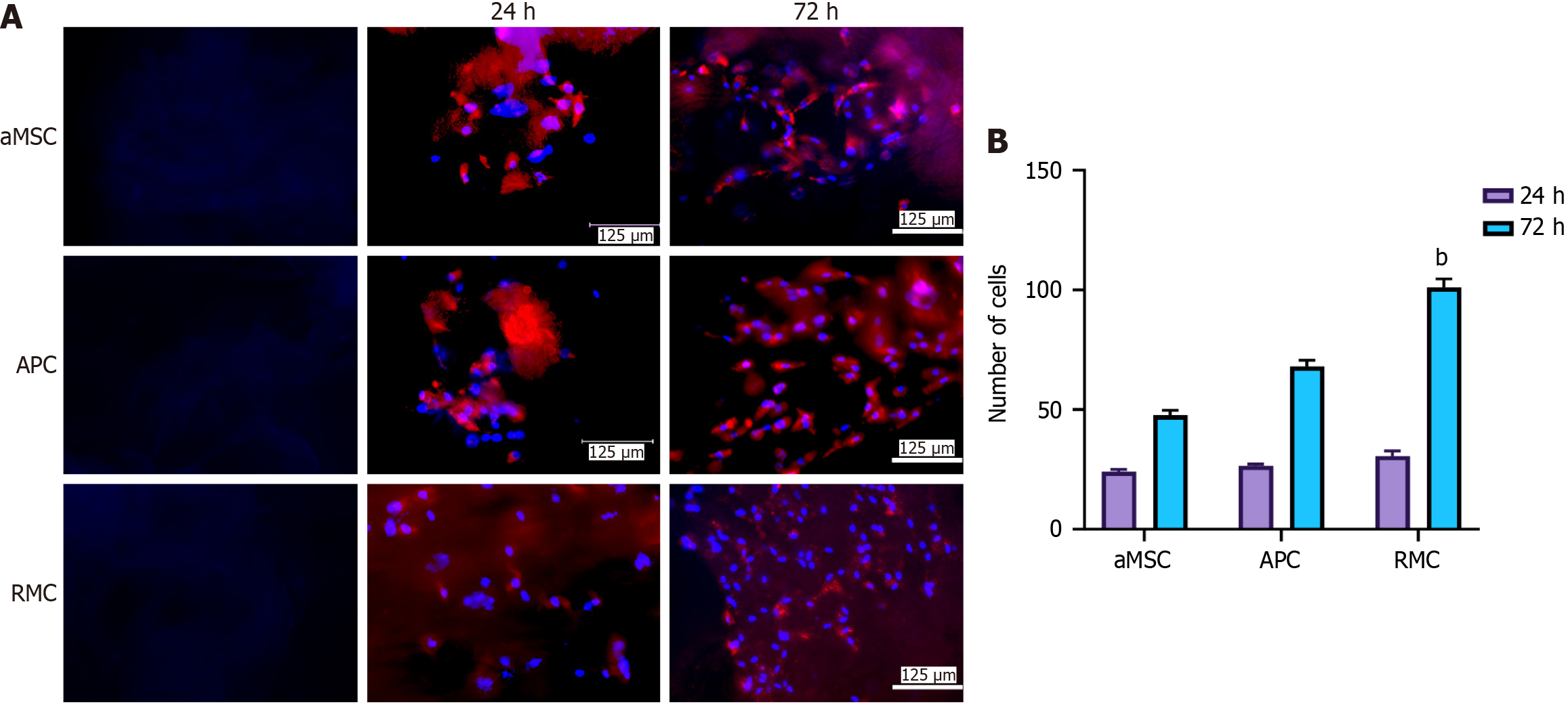
Figure 2 Effects of different extracellular matrix-sheets on cell attachment and proliferation in vitro.
A: Attachment and propagation of bone marrow mesenchymal stromal cells (bMSCs) on the fabricated adipocyte-derived MSC (aMSC-), antlerogenic periosteal cell (APC-), and antler reserve mesenchymal cell-extracellular matrix (RMC-ECM) sheets at two time points (24 and 72 h). 4’,6-diamidino-2-phenylindole (DAPI) staining: Blue and bMSCs red (pre-labelled with PKH26). First column: Decellularized ECM-sheets; second column: Cultured rat bMSCs 24 h after seeding on the sheets; third column: Cultured rat bMSCs 72 h after seeding on the sheets. Note that all three sheets were suitable for mesenchymal stromal cells to attach and proliferate; RMC-ECM-sheets not only had more cells but also less evidence of PKH26 label (red), indicating PKH26 color was more diluted due to subjecting more cycles of division; B: Quantification of cell numbers cultured on each ECM-sheet. Note that bMSCs were successfully attached and actively proliferated at 24 h after seeding, although no significant difference in cell numbers was detected amongst the three types of ECM-sheets. At 72 h, cell numbers of all groups were significantly increased compared to the corresponding group at 24 h (P < 0.001); and highly significantly differences in cell numbers were detected amongst these three groups: RMC group was higher (P < 0.01) than those of aMSC group and APC group; and APC group was significantly higher than that of aMSC group (P < 0.05). bMSCs: bone marrow mesenchymal stromal cell; aMSC: Adipocyte-derived mesenchymal stromal cell; APC: Antlerogenic periosteal cell; RMC: Antler reserve mesenchymal cell.
- Citation: Wang YS, Chu WH, Zhai JJ, Wang WY, He ZM, Zhao QM, Li CY. High quality repair of osteochondral defects in rats using the extracellular matrix of antler stem cells. World J Stem Cells 2024; 16(2): 176-190
- URL: https://www.wjgnet.com/1948-0210/full/v16/i2/176.htm
- DOI: https://dx.doi.org/10.4252/wjsc.v16.i2.176


