Copyright
©The Author(s) 2024.
World J Stem Cells. Feb 26, 2024; 16(2): 176-190
Published online Feb 26, 2024. doi: 10.4252/wjsc.v16.i2.176
Published online Feb 26, 2024. doi: 10.4252/wjsc.v16.i2.176
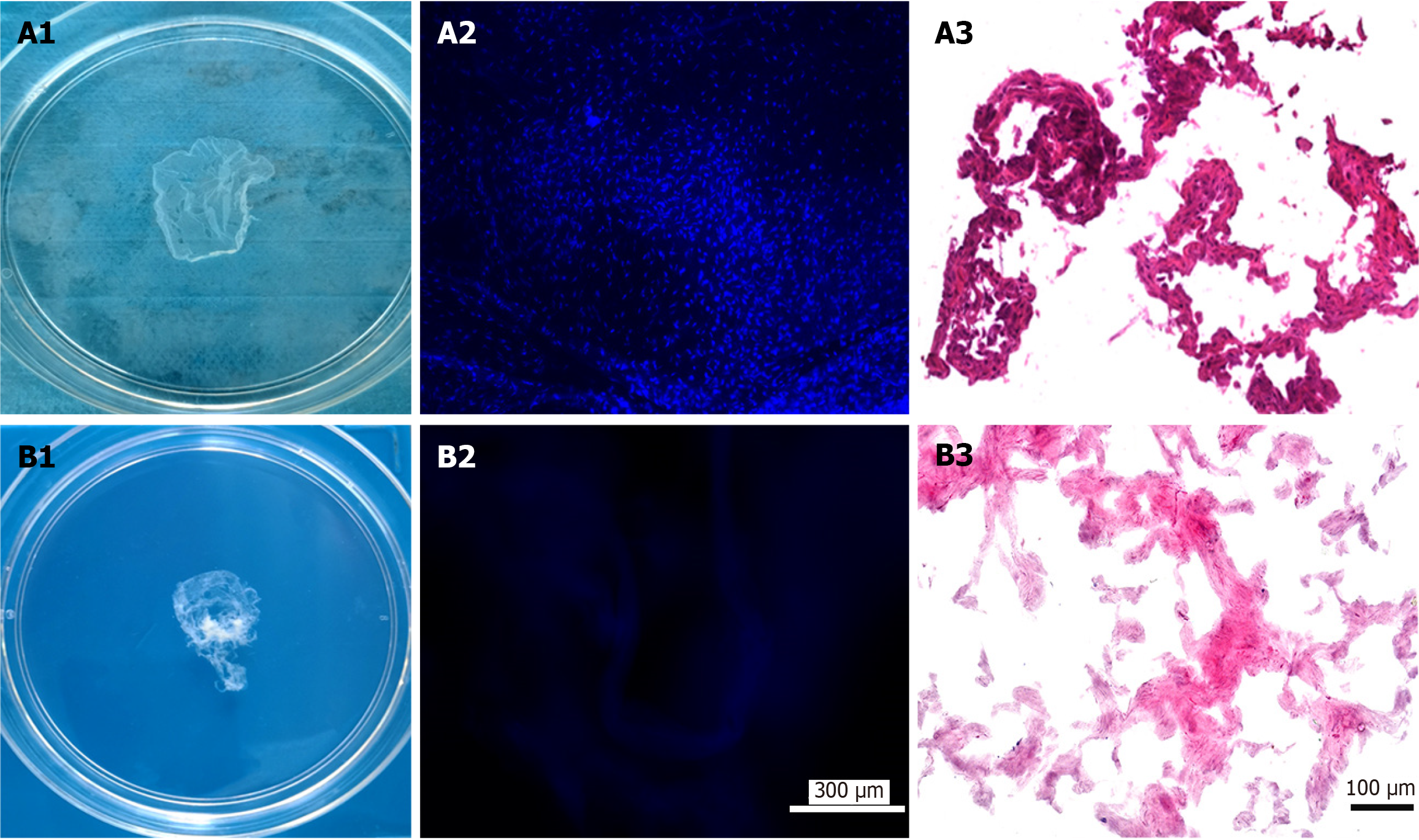
Figure 1 Fabrication of antler reserve mesenchymal cell-extracellular matrix-sheet.
A: Before decellularization: A1: Appearance of the sheet; A2: 4’,6-diamidino-2-phenylindole (DAPI) staining of the sheet; A3: Hematoxylin and eosin (HE) staining of the sheet; B: After decellularization: B1: Appearance of the sheet; B2: DAPI staining of the sheet; B3: HE staining of the sheet. Note that the decellularization process was successful and almost completely removed cells, evidenced by both DAPI and HE staining.
- Citation: Wang YS, Chu WH, Zhai JJ, Wang WY, He ZM, Zhao QM, Li CY. High quality repair of osteochondral defects in rats using the extracellular matrix of antler stem cells. World J Stem Cells 2024; 16(2): 176-190
- URL: https://www.wjgnet.com/1948-0210/full/v16/i2/176.htm
- DOI: https://dx.doi.org/10.4252/wjsc.v16.i2.176


