Copyright
©The Author(s) 2024.
World J Stem Cells. Dec 26, 2024; 16(12): 1047-1061
Published online Dec 26, 2024. doi: 10.4252/wjsc.v16.i12.1047
Published online Dec 26, 2024. doi: 10.4252/wjsc.v16.i12.1047
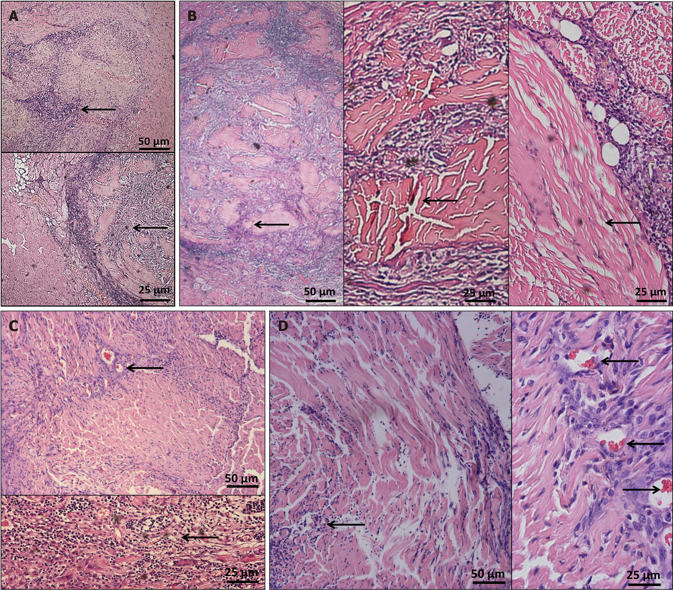
Figure 4 Hematoxylin and eosin staining after acellular scaffold implantation.
A: 3 days after surgery, a large number of inflammatory cells began to appear around the scaffold, including those surrounding the scaffold and spreading toward the middle; B: 7 days postoperatively, the number of inflammatory cells around the scaffold was further increased and the scaffold was further enveloped by the surrounding inflammatory cells; C: 3 weeks after surgery, the inflammatory cells around the scaffold began to fade; D: 6 weeks postoperatively, the inflammatory reaction further subsided and the shape of the scaffold was preserved.
- Citation: Qian C, Guo SY, Xu Z, Zhang ZQ, Li HD, Li H, Chen XS. Preliminary study on the preparation of lyophilized acellular nerve scaffold complexes from rabbit sciatic nerves with human umbilical cord mesenchymal stem cells. World J Stem Cells 2024; 16(12): 1047-1061
- URL: https://www.wjgnet.com/1948-0210/full/v16/i12/1047.htm
- DOI: https://dx.doi.org/10.4252/wjsc.v16.i12.1047


