Copyright
©The Author(s) 2024.
World J Stem Cells. Dec 26, 2024; 16(12): 1047-1061
Published online Dec 26, 2024. doi: 10.4252/wjsc.v16.i12.1047
Published online Dec 26, 2024. doi: 10.4252/wjsc.v16.i12.1047
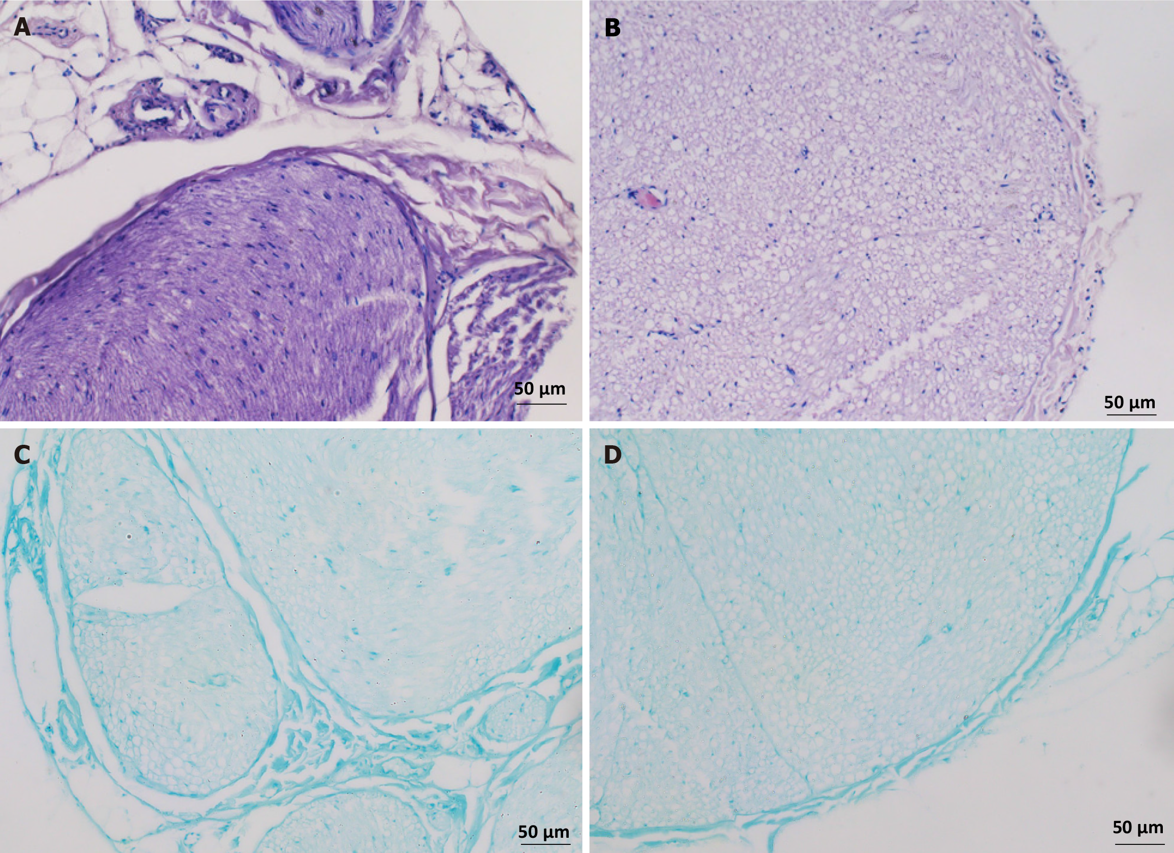
Figure 2 Histological observation of acellular scaffolds.
A: Hematoxylin and eosin (HE) staining shows the peripheral nerve fibers enveloped by the perineurium and a large amount of nuclear components may be seen in the peripheral nerves; B: After decellularization, the matrix structure of the HE-stained nerve fibers is preserved; however, the cell structure is no longer visible; C: Luxol fast blue (LFB) staining of peripheral nerve tissue reveals significant blue staining of myelin sheath tissue; D: LFB staining shows decreased staining of decellularized nerve tissue and myelin sheaths.
- Citation: Qian C, Guo SY, Xu Z, Zhang ZQ, Li HD, Li H, Chen XS. Preliminary study on the preparation of lyophilized acellular nerve scaffold complexes from rabbit sciatic nerves with human umbilical cord mesenchymal stem cells. World J Stem Cells 2024; 16(12): 1047-1061
- URL: https://www.wjgnet.com/1948-0210/full/v16/i12/1047.htm
- DOI: https://dx.doi.org/10.4252/wjsc.v16.i12.1047


