Copyright
©The Author(s) 2024.
World J Stem Cells. Nov 26, 2024; 16(11): 956-973
Published online Nov 26, 2024. doi: 10.4252/wjsc.v16.i11.956
Published online Nov 26, 2024. doi: 10.4252/wjsc.v16.i11.956
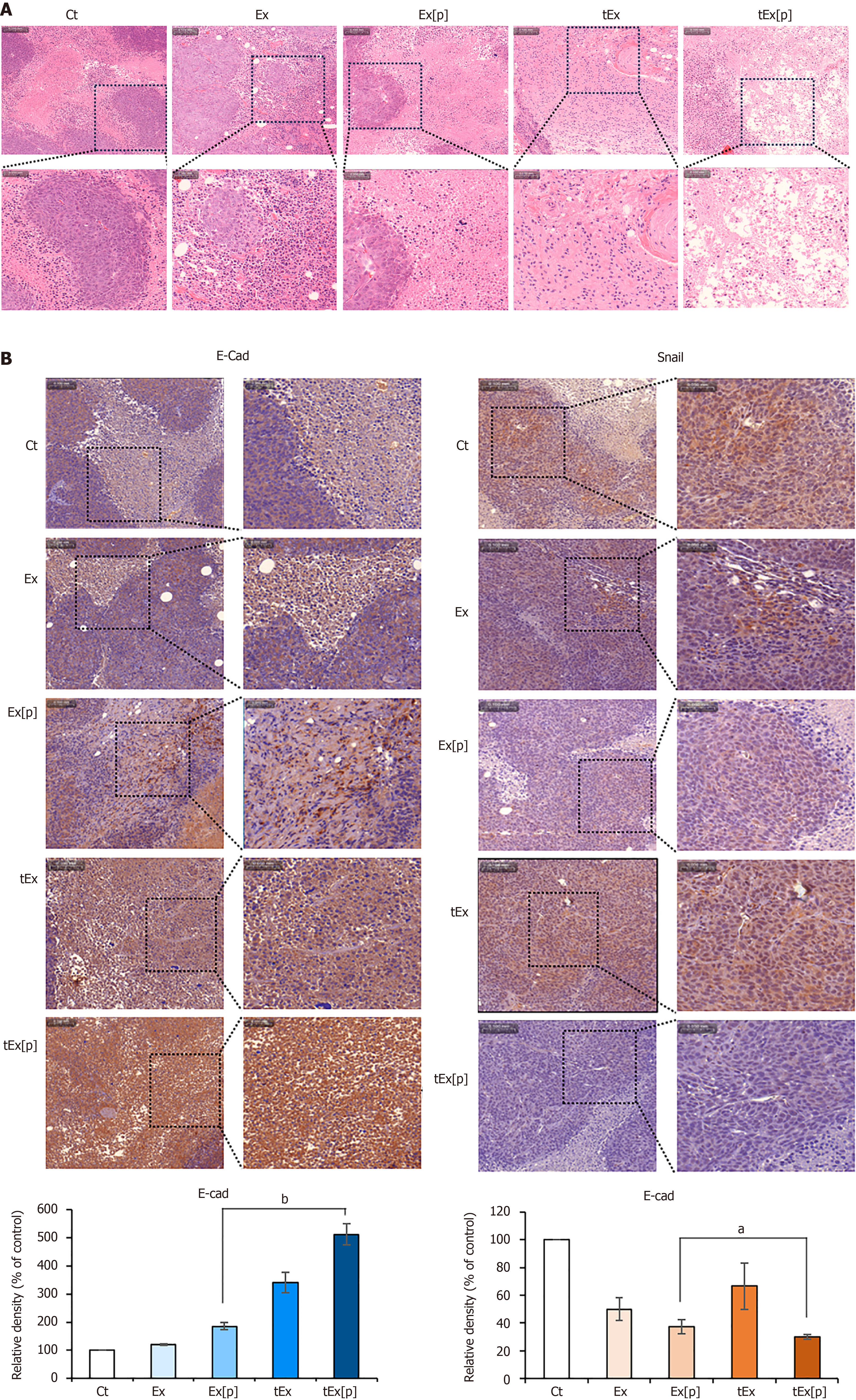
Figure 6 Histological analysis of excised tumor tissues in mouse colorectal cancer xenograft model.
A: Hematoxylin and eosin staining, showing that the small interfering peptidyl-prolyl cis-trans isomerase NIMA-interacting 1 RNA-loaded soluble a proliferation-inducing ligand-targeted exosome (tEx[p]) group exhibited a significantly reduced tumor cell density, indicating a pronounced effect on tumor growth suppression; B: Immunohistochemical analysis of epithelial-mesenchymal transition-related markers. The tEx[p] group showed increased immunoreactivity of the epithelial marker E-cadherin (left), while decreasing immunoreactivity of the mesenchymal marker Snail (right), underscoring its efficacy in inhibiting the epithelial-mesenchymal transition process in the colon cancer tissues. Values are presented as mean ± standard deviation of three independent experiments. Percentages of immunoreactive areas were measured using NIH ImageJ and expressed as relative values to those in control tissues. aP < 0.05, bP < 0.01. Ex: Exosome.
- Citation: Kim HJ, Lee DS, Park JH, Hong HE, Choi HJ, Kim OH, Kim SJ. Exosome-based strategy against colon cancer using small interfering RNA-loaded vesicles targeting soluble a proliferation-inducing ligand. World J Stem Cells 2024; 16(11): 956-973
- URL: https://www.wjgnet.com/1948-0210/full/v16/i11/956.htm
- DOI: https://dx.doi.org/10.4252/wjsc.v16.i11.956


