Copyright
©The Author(s) 2024.
World J Stem Cells. Nov 26, 2024; 16(11): 956-973
Published online Nov 26, 2024. doi: 10.4252/wjsc.v16.i11.956
Published online Nov 26, 2024. doi: 10.4252/wjsc.v16.i11.956
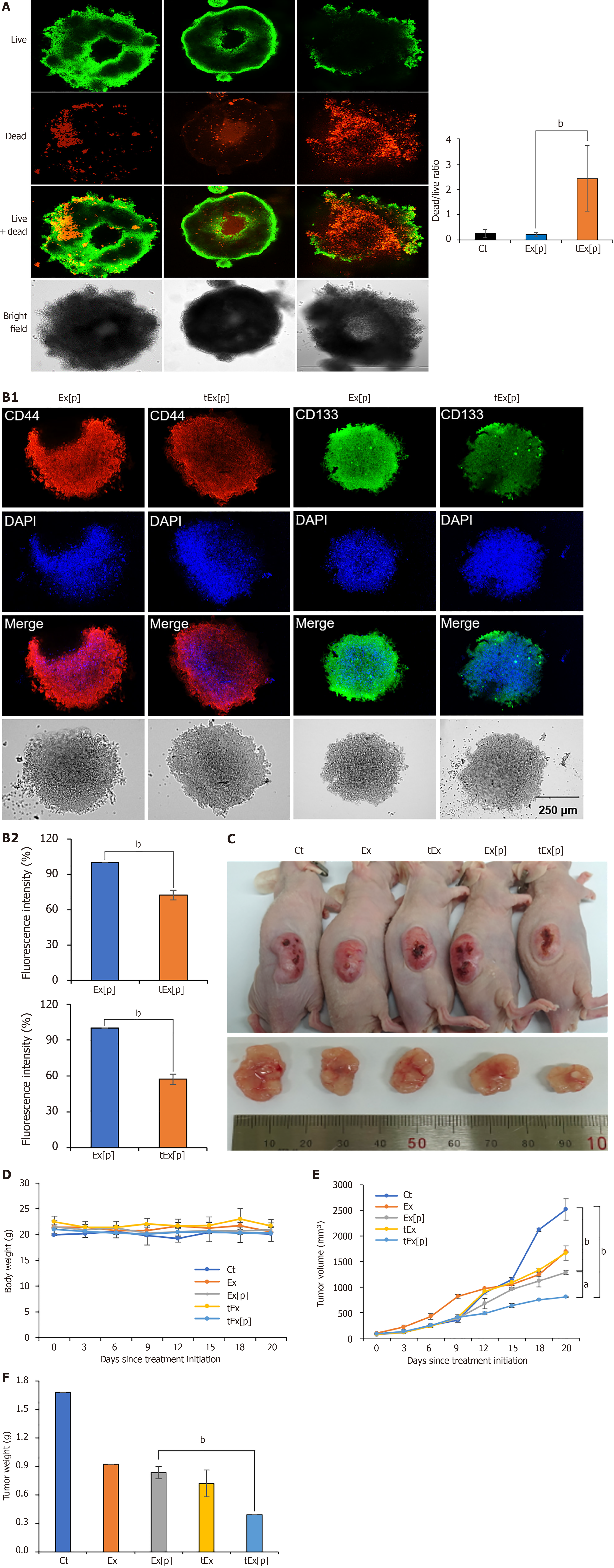
Figure 4 Spheroid-based and in vivo assessment of small interfering peptidyl-prolyl cis-trans isomerase NIMA-interacting 1 RNA-loaded soluble a proliferation-inducing ligand-targeted exosome efficacy in HCT116 models.
A: Effects of exosomes (Ex) loaded with siPIN1 (Ex[p]) and small interfering peptidyl-prolyl cis-trans isomerase NIMA-interacting 1 RNA-loaded soluble a proliferation-inducing ligand-targeted exosome (tEx[p]) on cell viability in HCT116-derived spheroids. Fluorescence microscopy images illustrate cell viability after treatments. Cells were stained using a LIVE/DEAD staining kit to distinguish live cells (green) from dead cells (red) (left panel). Representative images of spheroids treated with negative control, Ex[p], and tEx[p] (right panel). Quantification of the dead/live ratio, showing a significant increase in cell death in the tEx[p] treated spheroids compared to Ex[p] treated ones. This increase suggests that tEx[p] could also affect the viability of cancer stem cells within the spheroids, indicating its potential efficacy against tumor resilience. Error bars denote standard deviation based on three independent experiments; B: Immunofluorescence analysis of cancer stem cell markers CD44 and CD133 in spheroids treated with Ex[p] and tEx[p] for 24 hours. The expression of CD44 (red) and CD133 (green) is reduced in tEx[p] treated spheroids compared to Ex[p] treated spheroids, indicating a decrease in cancer stem cell populations. Nuclei are counterstained with DAPI (blue). Images are representative of three independent experiments; C: Xenograft appearance and size comparison in each group; D: Comparision of body weight measurements, demonstrating no significant weight differences among the groups, suggesting minimal systemic toxicity; E: Measurements of tumor volumes. The tEx[p] group demonstrated the smallest tumor volumes in the comparison of tumor sizes; F: Measurements of tumor volumes on day 20 post-treatment. The tEx[p] group exhibited significantly reduced tumor weights compared to other groups in the tumor weight comparison. aP < 0.05, bP < 0.01.
- Citation: Kim HJ, Lee DS, Park JH, Hong HE, Choi HJ, Kim OH, Kim SJ. Exosome-based strategy against colon cancer using small interfering RNA-loaded vesicles targeting soluble a proliferation-inducing ligand. World J Stem Cells 2024; 16(11): 956-973
- URL: https://www.wjgnet.com/1948-0210/full/v16/i11/956.htm
- DOI: https://dx.doi.org/10.4252/wjsc.v16.i11.956


