Copyright
©The Author(s) 2024.
World J Stem Cells. Nov 26, 2024; 16(11): 956-973
Published online Nov 26, 2024. doi: 10.4252/wjsc.v16.i11.956
Published online Nov 26, 2024. doi: 10.4252/wjsc.v16.i11.956
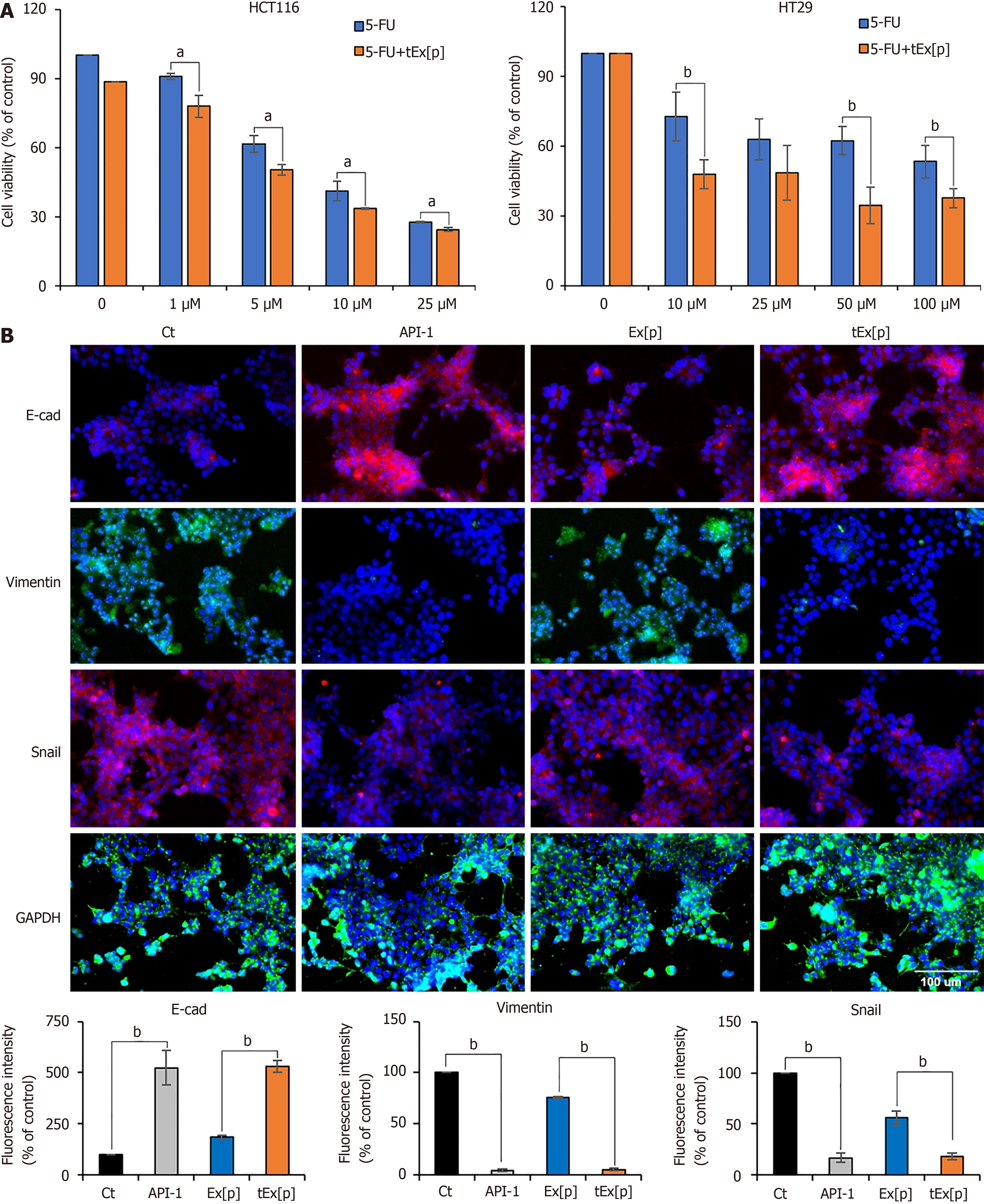
Figure 3 Enhancing chemosensitivity and inhibiting epithelial-mesenchymal transition in colon cancer cell lines using small interfering peptidyl-prolyl cis-trans isomerase NIMA-interacting 1 RNA-loaded exosomes.
A: Cell viability assay under increasing concentrations of 5-fluorouridine (5-FU) alone and in combination with small interfering peptidyl-prolyl cis-trans isomerase NIMA-interacting 1 RNA (siPIN1)-loaded soluble a proliferation-inducing ligand-targeted exosome (tEx[p]) in HCT116 (left) and HT29 (right) colon cancer cell lines. This figure illustrates the cell viability expressed as a percentage relative to untreated controls in two distinct colon cancer cell lines subjected to escalating doses of 5-FU, both alone and in conjunction with tEx[p]. The data highlight that the concomitant use of tEx[p] with 5-FU could enhance the chemotherapeutic response by further diminishing cancer cell survival; B: Immunofluorescence of epithelial-mesenchymal transition (EMT) markers upon treatment of API-1 (PIN inhibitor) and siPIN encapsulated exosomes (Ex). Immunofluorescence of EMT markers was performed for the determination of the effects of API-1, Ex(siPIN1), and tEx(siPIN1) on EMT in HCT116 cells. Treatment of API-1 (20 μM) and tEx(siPIN1; 100 pmol) effectively inhibited EMT, as demonstrated by the highest increase in E-cadherin expression and the lowest decrease in Snail and Vimentin expression among all treatment groups. Percentages of immunoreactive areas were measured using NIH ImageJ and expressed as relative values to those in control cells. Values are presented as mean ± standard deviation (SD) of three independent experiments. Values are presented as mean ± SD of three independent experiments. aP < 0.05, bP < 0.01.
- Citation: Kim HJ, Lee DS, Park JH, Hong HE, Choi HJ, Kim OH, Kim SJ. Exosome-based strategy against colon cancer using small interfering RNA-loaded vesicles targeting soluble a proliferation-inducing ligand. World J Stem Cells 2024; 16(11): 956-973
- URL: https://www.wjgnet.com/1948-0210/full/v16/i11/956.htm
- DOI: https://dx.doi.org/10.4252/wjsc.v16.i11.956


