Copyright
©The Author(s) 2023.
World J Stem Cells. Sep 26, 2023; 15(9): 931-946
Published online Sep 26, 2023. doi: 10.4252/wjsc.v15.i9.931
Published online Sep 26, 2023. doi: 10.4252/wjsc.v15.i9.931
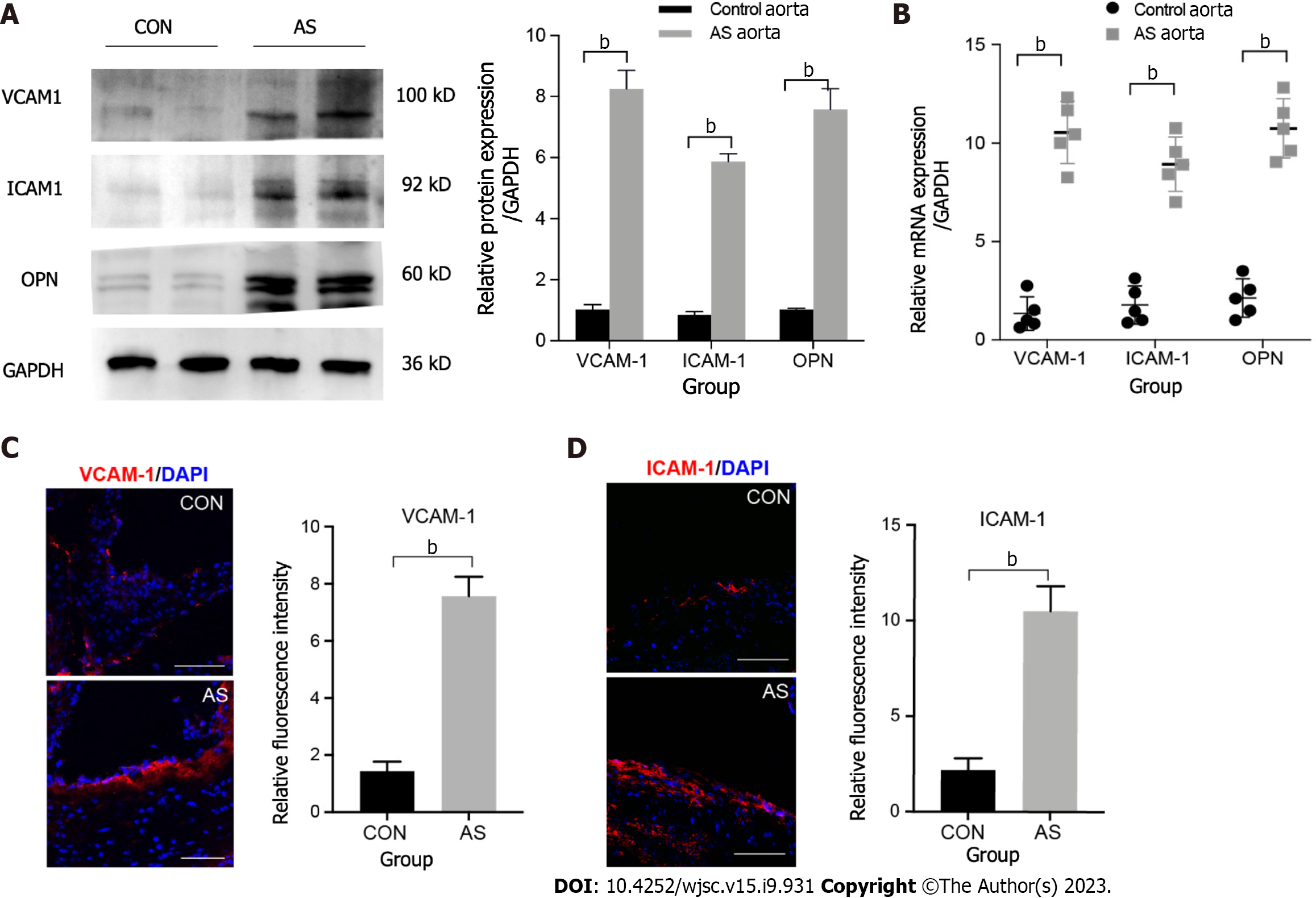
Figure 1 Expression of inflammatory factors in the vascular atherosclerotic plaque.
A: Expression of mouse vascular cell adhesion molecule-1 (VCAM-1), intercellular cell adhesion molecule-1 (ICAM-1), and osteopontin (OPN) expression in total tissue lysates of normal and atherosclerotic (AS) aorta analyzed using western blot. GAPDH was used as the internal control. The experiment was repeated thrice with tissues isolated from independent mice; a representative blot is shown; B: Expression levels of various inflammatory factors involved in atherosclerosis analyzed using quantitative real-time polymerase chain reaction of mRNA samples extracted from normal and AS vessels of three independent mice. Data are presented as the mean ± SEM for each group. Fold change represents the expression of each inflammatory factor in AS vessel of a mice fed with high fat diet for 12 wk compared with that in normal blood vessel; C: Representative images of normal and AS vascular sections stained for VCAM1 (red) and ICAM1 (red). The experiment was repeated three times with tissues isolated from independent mice; a representative image is shown. Nuclei were visualized by DAPI staining (blue). Scale bars = 100 mm; D: Representative images of normal and AS vascular sections stained for ICAM1 (red). The experiment was repeated three times with tissues isolated from independent mice; a representative image is shown. Nuclei were visualized by DAPI staining (blue). Scale bars = 100 μm. bP < 0.001. AS: Atherosclerotic; OPN: Osteopontin; qRT-PCR: Quantitative real-time polymerase chain reaction; SEM: Standard error of the mean; VCAM-1: Vascular cell adhesion molecule-1; ICAM-1: Intercellular cell adhesion molecule-1.
- Citation: Hu HJ, Xiao XR, Li T, Liu DM, Geng X, Han M, Cui W. Integrin beta 3-overexpressing mesenchymal stromal cells display enhanced homing and can reduce atherosclerotic plaque. World J Stem Cells 2023; 15(9): 931-946
- URL: https://www.wjgnet.com/1948-0210/full/v15/i9/931.htm
- DOI: https://dx.doi.org/10.4252/wjsc.v15.i9.931


