Copyright
©The Author(s) 2023.
World J Stem Cells. Sep 26, 2023; 15(9): 876-896
Published online Sep 26, 2023. doi: 10.4252/wjsc.v15.i9.876
Published online Sep 26, 2023. doi: 10.4252/wjsc.v15.i9.876
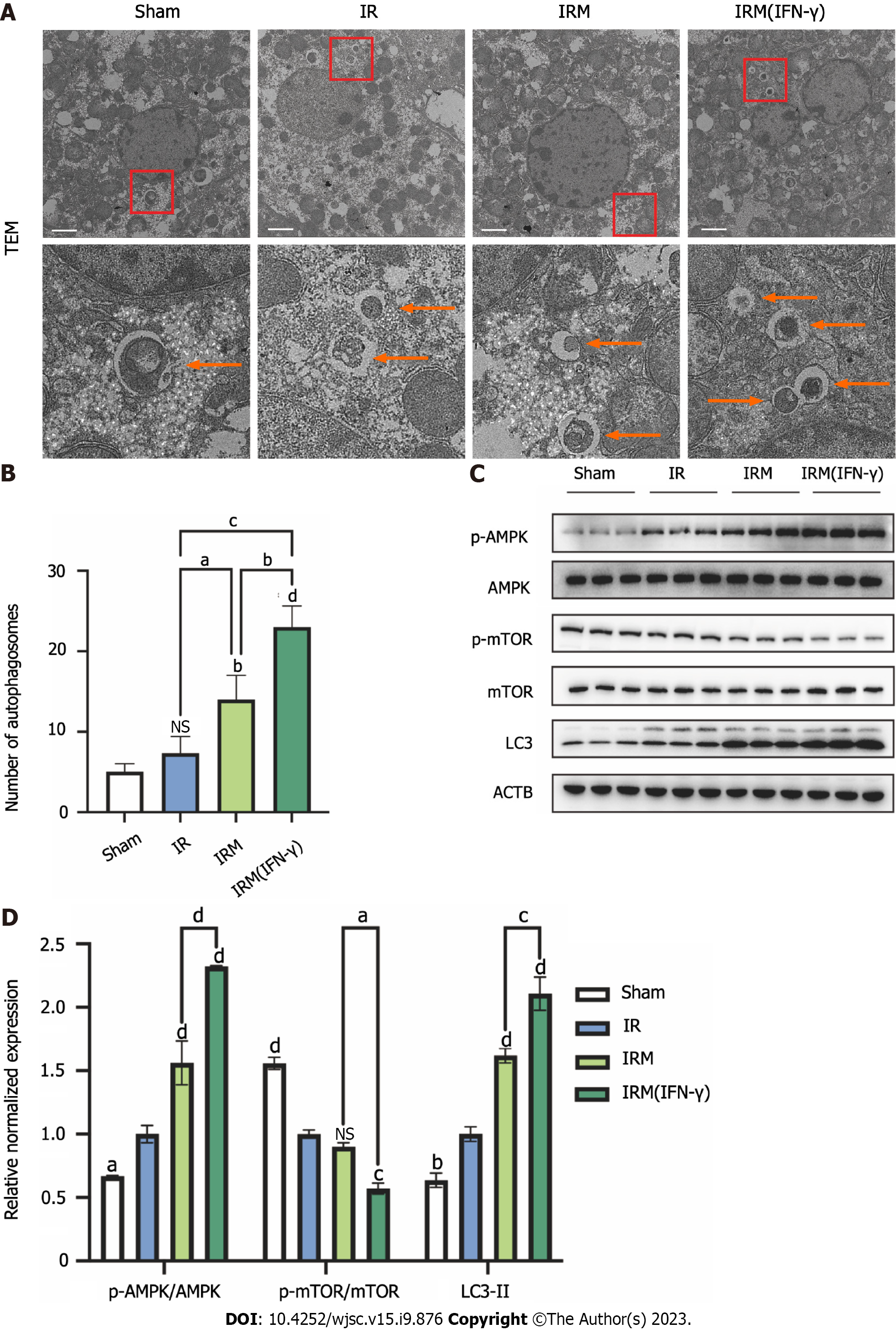
Figure 6 Interferon-γ-primed menstrual blood-derived stromal cells enhanced autophagy in mouse livers.
A: Representative TEM images were used to determine the number of autophagic vacuoles in liver tissues from each experimental group. Scale bar 2 μm. The images in the second row are partial magnifications of the regions indicated by the red box; B: The number of autophagosomes was counted by TEM. The data are expressed as the means ± SEMs (n = 3/group); C: The expression of p-AMPK, AMPK, p-mammalian target of rapamycin (p-mTOR), mTOR, LC3, and ACTB in liver tissues was determined by western blotting; D: The relative normalized expression was quantified based on the intensities of the western blot bands (n = 3/group). The data are presented as the means ± SEMs. aP < 0.05, bP < 0.01, cP < 0.001, dP < 0.0001. NS represents not statistically significant. All P values were obtained by one-way ANOVA. MenSCs: Menstrual blood-derived stromal cells; IFN-γ: Interferon-γ; IR: Ischemia-reperfusion; IRM: Combination treated with ischemia-reperfusion and menstrual blood-derived stromal cells; IRM (IFN-γ): Combination treated with ischemia-reperfusion and interferon-γ-primed menstrual blood-derived stromal cells; TEM: Transmission electron microscopy; AMPK: The AMP-activated protein kinase signaling pathway; mTOR: Mammalian target of rapamycin; Tregs: Regulatory T cells.
- Citation: Zhang Q, Zhou SN, Fu JM, Chen LJ, Fang YX, Xu ZY, Xu HK, Yuan Y, Huang YQ, Zhang N, Li YF, Xiang C. Interferon-γ priming enhances the therapeutic effects of menstrual blood-derived stromal cells in a mouse liver ischemia-reperfusion model. World J Stem Cells 2023; 15(9): 876-896
- URL: https://www.wjgnet.com/1948-0210/full/v15/i9/876.htm
- DOI: https://dx.doi.org/10.4252/wjsc.v15.i9.876


