Copyright
©The Author(s) 2023.
World J Stem Cells. Sep 26, 2023; 15(9): 876-896
Published online Sep 26, 2023. doi: 10.4252/wjsc.v15.i9.876
Published online Sep 26, 2023. doi: 10.4252/wjsc.v15.i9.876
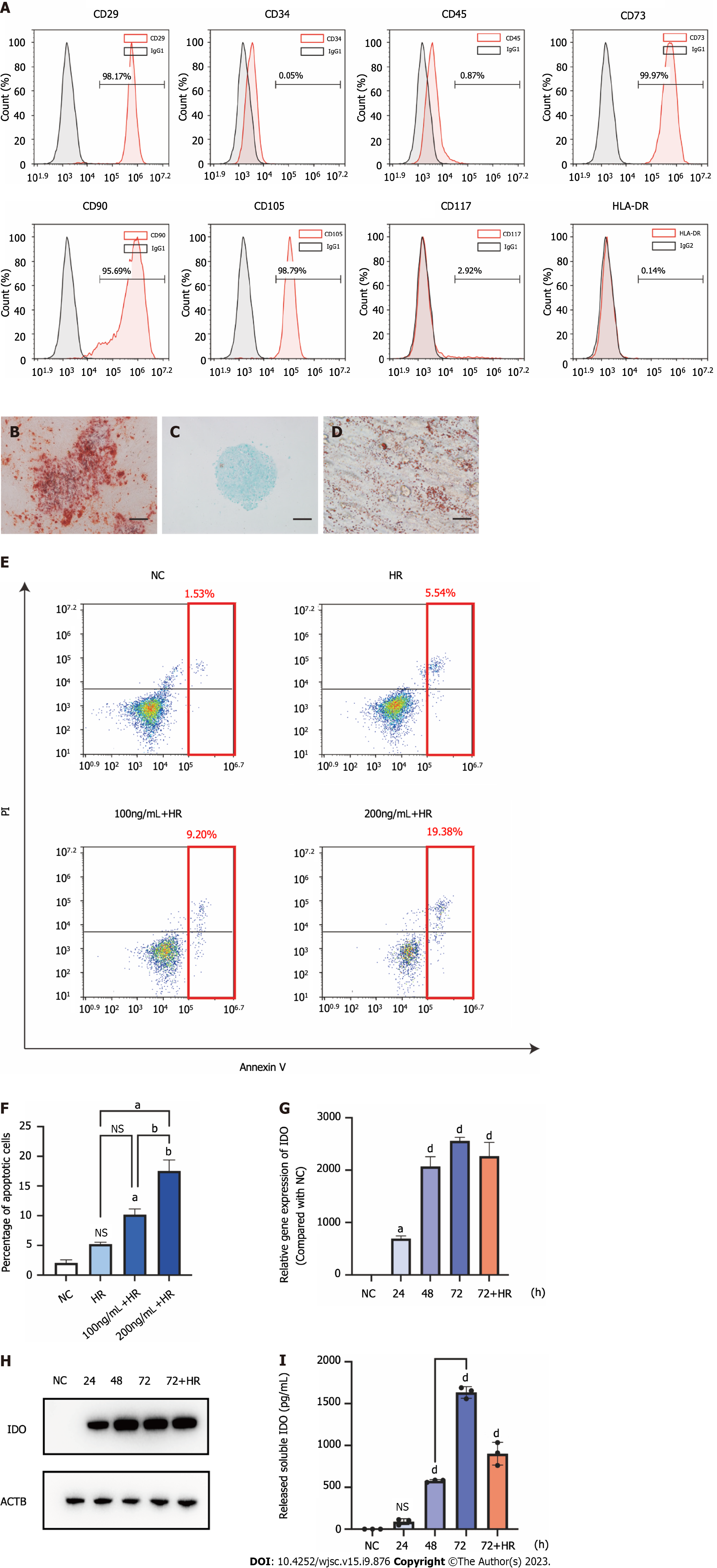
Figure 2 Characterization of menstrual blood-derived stromal cells and menstrual blood-derived stromal cells primed with interferon-γ.
A: The surface marker expression of menstrual blood-derived stromal cells (MenSCs) was assessed by flow cytometry analysis; B: The osteogenic differentiation potential of MenSCs. Scale bar 200 μm; C: The chondrogenic differentiation potential of MenSCs. Scale bar 200 μm; D: The adipogenic differentiation potential of MenSCs. Scale bar 200 μm; E: MenSC apoptosis was detected by flow cytometry. The red box indicates the sum of late and early apoptotic cells, which represent the overall level of apoptosis; F: The overall level of apoptosis was analyzed; G: After MenSC priming with 100 ng/mL interferon-γ for 24 h, 48 h, 72 h, and 72 h + hypoxia/reoxygenation, the mRNA levels of indoleamine 2,3-dioxygenase (IDO) in MenSCs were determined by quantitative real-time reverse transcription polymerase chain reaction analysis; H: The protein level of IDO in MenSCs was determined by western blotting; I: The level of IDO secreted by MenSCs into the cell culture supernatant was measured by ELISA. The data are shown as the means ± SEMs, n = 3. aP < 0.05, bP < 0.01, cP < 0.001, dP < 0.0001. NS represents not statistically significant. All P values were obtained by one-way ANOVA. HLA-DR: Human leukocyte antigen-DR; IgG: Immunoglobulin G; NC: Negative control; HR: Hypoxia/reoxygenation; PI: Propidium iodide; IDO: Indoleamine 2,3-dioxygenase.
- Citation: Zhang Q, Zhou SN, Fu JM, Chen LJ, Fang YX, Xu ZY, Xu HK, Yuan Y, Huang YQ, Zhang N, Li YF, Xiang C. Interferon-γ priming enhances the therapeutic effects of menstrual blood-derived stromal cells in a mouse liver ischemia-reperfusion model. World J Stem Cells 2023; 15(9): 876-896
- URL: https://www.wjgnet.com/1948-0210/full/v15/i9/876.htm
- DOI: https://dx.doi.org/10.4252/wjsc.v15.i9.876


