Copyright
©The Author(s) 2023.
World J Stem Cells. Aug 26, 2023; 15(8): 842-865
Published online Aug 26, 2023. doi: 10.4252/wjsc.v15.i8.842
Published online Aug 26, 2023. doi: 10.4252/wjsc.v15.i8.842
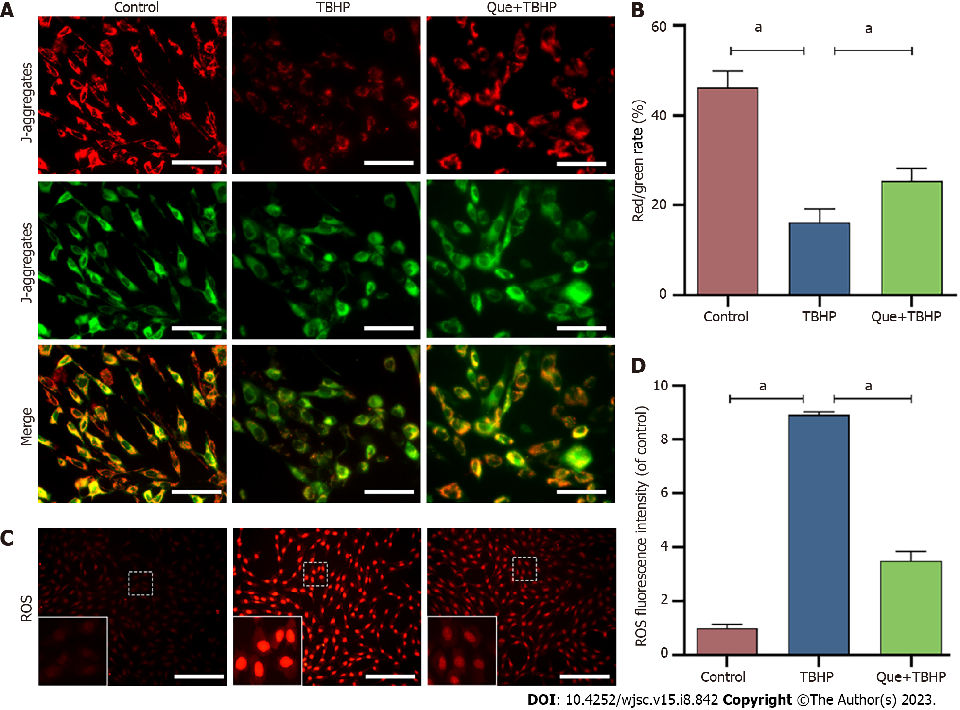
Figure 4 Mitochondrial membrane potential assay and reactive oxygen species assay.
A: Results of Mitochondrial membrane potential (MMP) in different groups detected by fluorescence. Red fluorescence represents the mitochondrial aggregate JC-1 and green fluorescence indicates the monomeric JC-1. Scale bar = 100 μm; B: Quantitative analysis of MMP results; C: Results of ROS in different groups detected by fluorescence. Red fluorescence represents high level of reactive oxygen species assay (ROS). Scale bar = 200 μm; D: Quantitative analysis of ROS results. Data are represented as mean ± SD. Significant differences between groups are indicated as aP < 0.05, n = 3. TBHP: Tert-butyl hydroperoxide; Que: Quercetin; ROS: Reactive oxygen species assay.
- Citation: Zhao WJ, Liu X, Hu M, Zhang Y, Shi PZ, Wang JW, Lu XH, Cheng XF, Tao YP, Feng XM, Wang YX, Zhang L. Quercetin ameliorates oxidative stress-induced senescence in rat nucleus pulposus-derived mesenchymal stem cells via the miR-34a-5p/SIRT1 axis. World J Stem Cells 2023; 15(8): 842-865
- URL: https://www.wjgnet.com/1948-0210/full/v15/i8/842.htm
- DOI: https://dx.doi.org/10.4252/wjsc.v15.i8.842


