Copyright
©The Author(s) 2023.
World J Stem Cells. Aug 26, 2023; 15(8): 821-841
Published online Aug 26, 2023. doi: 10.4252/wjsc.v15.i8.821
Published online Aug 26, 2023. doi: 10.4252/wjsc.v15.i8.821
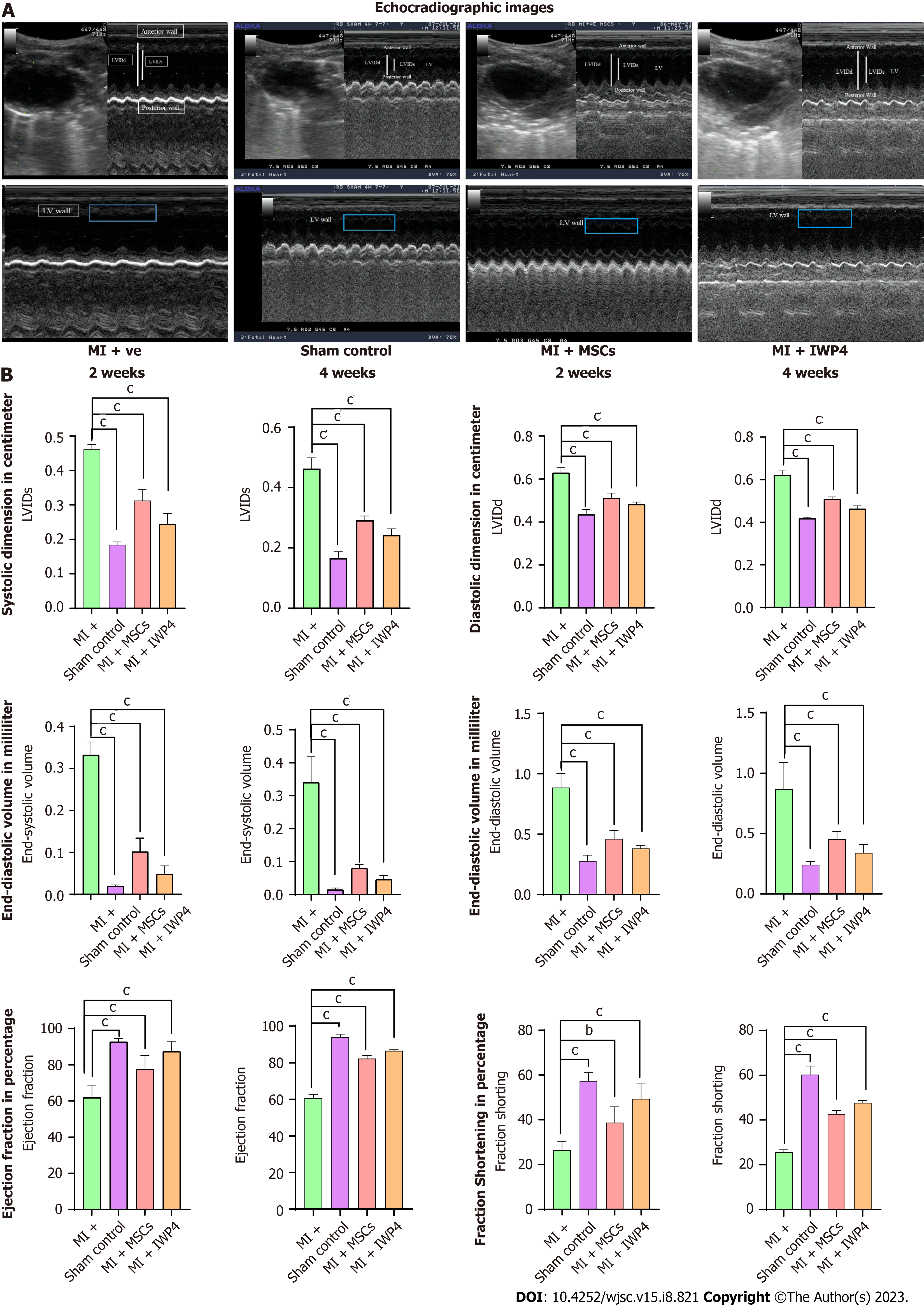
Figure 6 Cardiac functional analysis by echocardiography.
A: Ultrasound images taken at parasternal long axis showing B and M mode scans of left ventricle in sham control, myocardial infarction (MI+) group, mesenchymal stem cells (MI+MSC) treated group, and inhibitor Wnt production-4 (IWP-4) (MI+IWP-4) treated groups; B: Bar graphs representing cardiac functional analysis in terms of left ventricular systolic internal dimensions, left ventricular diastolic internal dimensions, end-systolic volume and end-diastolic volume, ejection fraction and fractional shortening, after 2 and 4 wk of MI model development. Both untreated and IWP-4 treated MSC groups showed cardiac functional improvement. However, IWP-4 treated group exhibited more significant results. Statistical analysis was performed using One-way ANOVA followed by Bonferroni post-hoc test. Data are presented as mean ± SEM with significance level P < 0.05 (where bP < 0.01; cP < 0.001). IWP-4: Inhibitor Wnt production-4; MSC: Mesenchymal stem cell; MI: Myocardial infarction; LVIDs: Left ventricular systolic internal dimensions; LVIDd: Left ventricular diastolic internal dimensions.
- Citation: Muneer R, Qazi REM, Fatima A, Ahmad W, Salim A, Dini L, Khan I. Wnt signaling pathway inhibitor promotes mesenchymal stem cells differentiation into cardiac progenitor cells in vitro and improves cardiomyopathy in vivo. World J Stem Cells 2023; 15(8): 821-841
- URL: https://www.wjgnet.com/1948-0210/full/v15/i8/821.htm
- DOI: https://dx.doi.org/10.4252/wjsc.v15.i8.821


