Copyright
©The Author(s) 2023.
World J Stem Cells. Jul 26, 2023; 15(7): 768-780
Published online Jul 26, 2023. doi: 10.4252/wjsc.v15.i7.768
Published online Jul 26, 2023. doi: 10.4252/wjsc.v15.i7.768
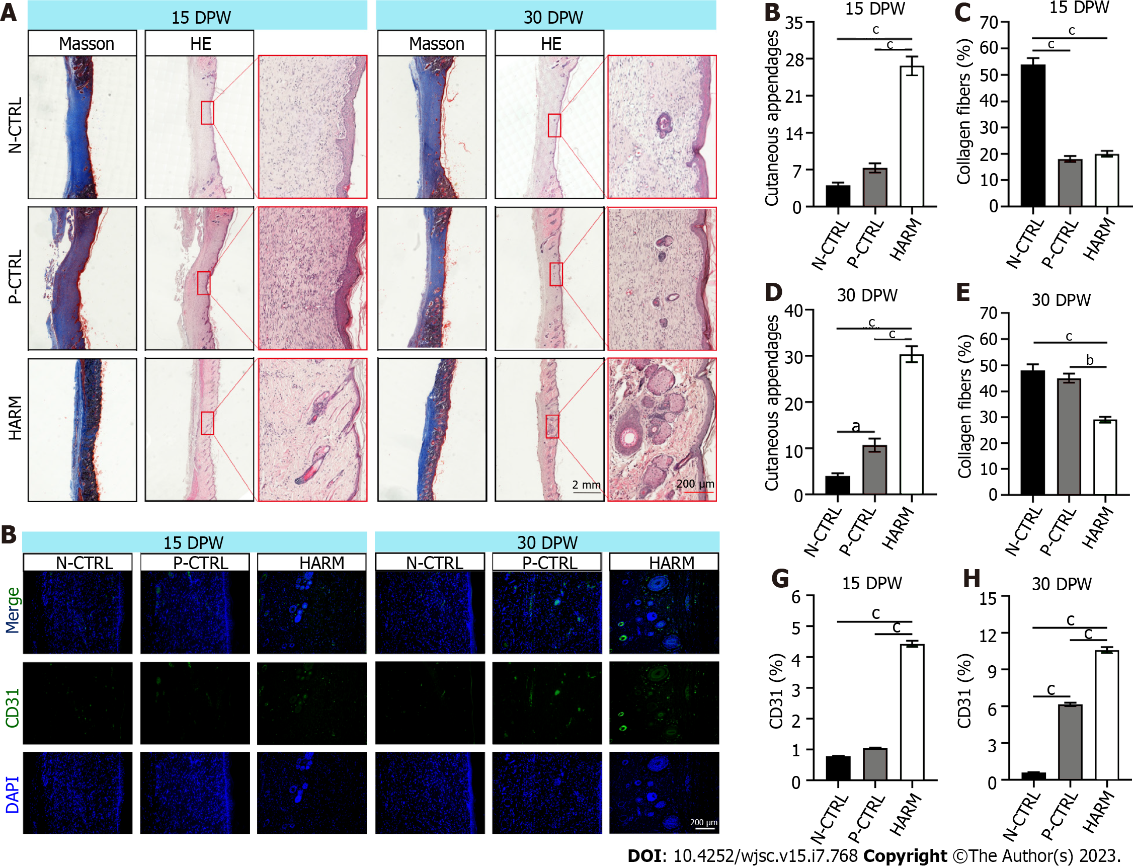
Figure 3 Effects of hydrogels from the antler reserve mesenchyme on the quality of wound healing.
A: Histological sections of the healed skin stained with HE and Masson; B-E: Quantification of cutaneous appendages and the collagen fiber accounts (blue area in Masson staining); F: CD31 immunofluorescence staining; G and H: Expression percentage of CD31. Note that the highest number of cutaneous appendages, least account of collagen fibers, and highest percent were found in the HARM group in the healed tissues compared to the N-CTRL and P-CTRL groups. DPW: Days post-wounding; HARM: Hydrogels derived from antler reserve mesenchyme; N-CTRL: Negative control; P-CTRL: Positive control. n = 3; mean ± SEM; statistical significance set at aP < 0.05, bP < 0.01, cP < 0.001.
- Citation: Zhang GK, Ren J, Li JP, Wang DX, Wang SN, Shi LY, Li CY. Injectable hydrogel made from antler mesenchyme matrix for regenerative wound healing via creating a fetal-like niche. World J Stem Cells 2023; 15(7): 768-780
- URL: https://www.wjgnet.com/1948-0210/full/v15/i7/768.htm
- DOI: https://dx.doi.org/10.4252/wjsc.v15.i7.768


