Copyright
©The Author(s) 2023.
World J Stem Cells. Jul 26, 2023; 15(7): 768-780
Published online Jul 26, 2023. doi: 10.4252/wjsc.v15.i7.768
Published online Jul 26, 2023. doi: 10.4252/wjsc.v15.i7.768
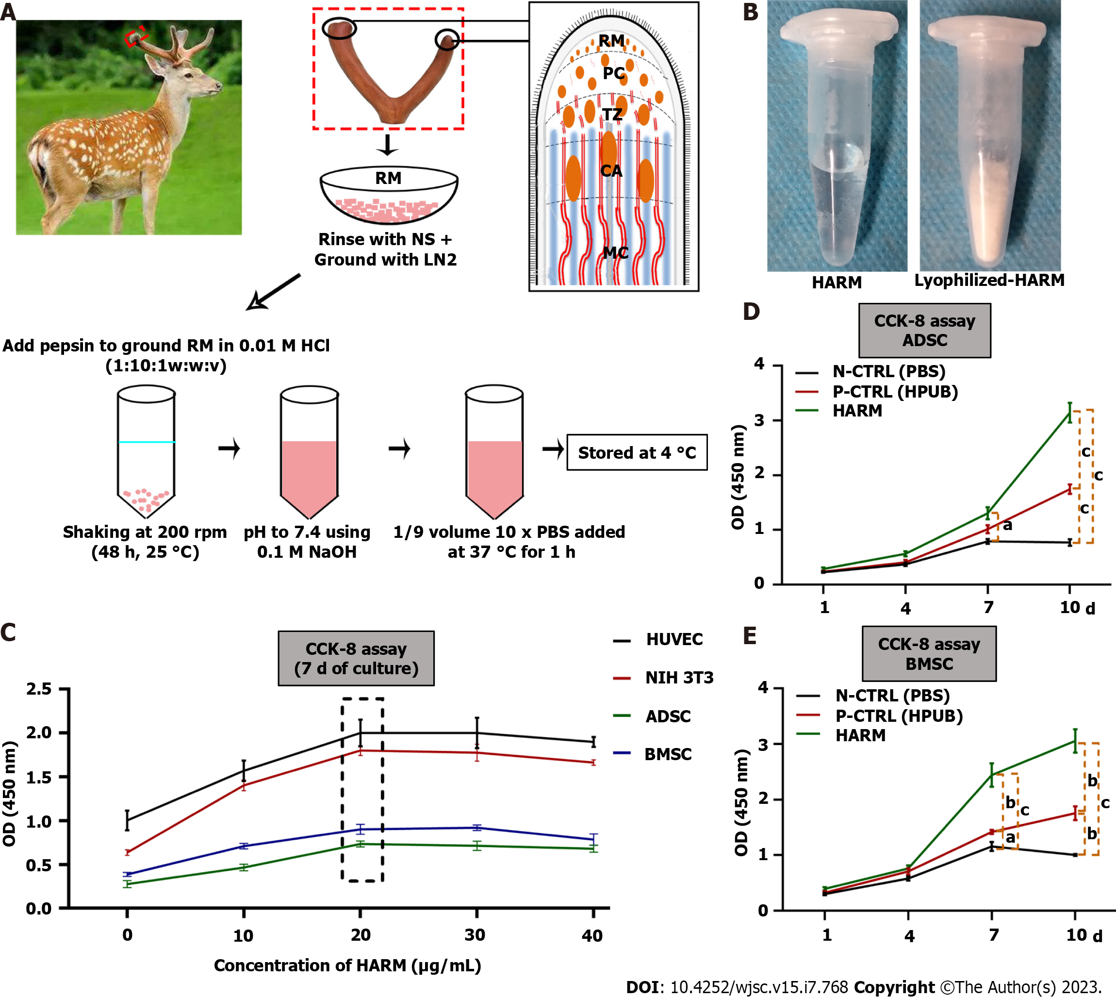
Figure 1 Preparation of hydrogels from the antler reserve mesenchyme matrix and its effects on cell proliferation invitro.
A: Location of reserve mesenchyme (RM) and fabrication of hydrogels from the antler reserve mesenchyme matrix (HARM); B: HARM (top) and lyophilized-HARM (bottom); C: Screening of the optimal concentration of HARM on cell [HUVEC, NIH 3T3, adipose mesenchymal stem cells (ADSC), and bone marrow mesenchymal stem cells (BMSC)] proliferation rate; D and E: Comparison of HARM with hydrogels from porcine urinary bladder matrix (HPUB) on cell (ADSC and BMSC) proliferation rate. Note that the concentration of 20 μg/mL HARM had the strongest stimulation over the others on proliferation rate of HUVEC, NIH 3T3, ADSC, and BMSC; moreover, proliferation rates of ADSC and BMSC in HARM were stronger than in N-CTRL (PBS) or in P-CTRL (HPUB). RM: Reserve mesenchyme; PC: Pre-cartilage; TZ: Transitional zone; CA: Cartilage; NS: Normal saline; LN2: Liquid nitrogen; HARM: Hydrogels derived from antler reserve mesenchyme; HPUB: Hydrogels from porcine urinary bladder matrix; HUVEC: Human umbilical vein endothelial cells; NIH 3T3: Mouse embryonic fibroblasts; ADSC: Adipose mesenchymal stem cells; BMSC: Bone marrow mesenchymal stem cells; N-CTRL: Negative control; P-CTRL: Positive control. n = 3; mean ± SEM; statistically significance set at aP < 0.05, bP < 0.01, and cP < 0.001.
- Citation: Zhang GK, Ren J, Li JP, Wang DX, Wang SN, Shi LY, Li CY. Injectable hydrogel made from antler mesenchyme matrix for regenerative wound healing via creating a fetal-like niche. World J Stem Cells 2023; 15(7): 768-780
- URL: https://www.wjgnet.com/1948-0210/full/v15/i7/768.htm
- DOI: https://dx.doi.org/10.4252/wjsc.v15.i7.768


