Copyright
©The Author(s) 2023.
World J Stem Cells. Jul 26, 2023; 15(7): 751-767
Published online Jul 26, 2023. doi: 10.4252/wjsc.v15.i7.751
Published online Jul 26, 2023. doi: 10.4252/wjsc.v15.i7.751
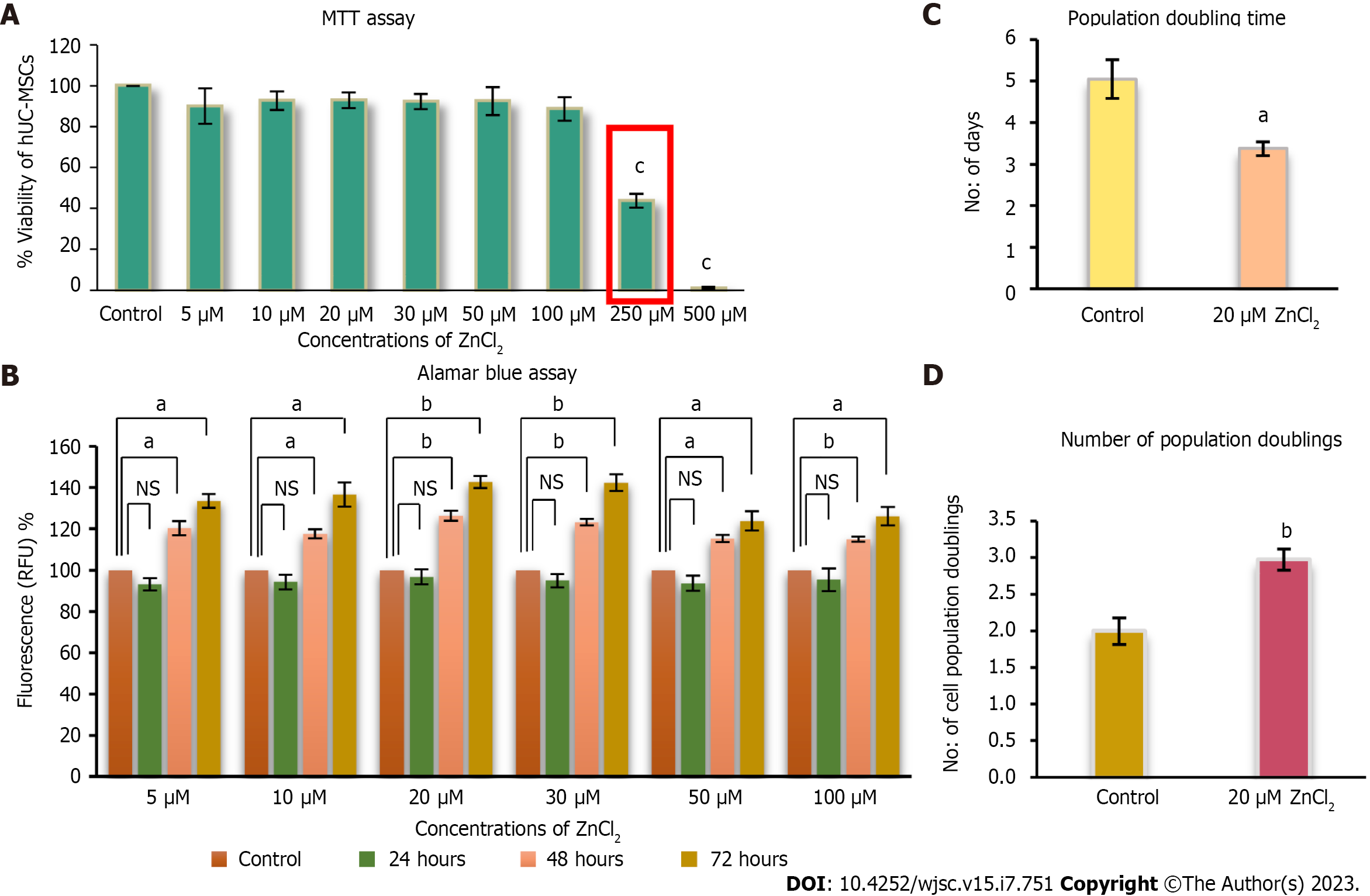
Figure 3 Cytotoxic analysis of ZnCl2 for human umbilical cord-derived mesenchymal stem cells.
A: Concentrations of ZnCl2 5 µM, 10 µM, 20 µM, 30 µM, 50 µM, 100 µM, 250 µM, and 500 µM, were analyzed for their cytotoxicity by MTT assay. Concentrations from 5 µM to 100 µM did not show any significant decrease in viability (Percent) of human umbilical cord (hUC)-derived mesenchymal stem cells (MSCs) compared to control. 250 µM reflected IC50 value of ZnCl2, and both 250 µM and 500 µM concentrations were found to be significantly (cP ≤ 0.001) cytotoxic. Results were analyzed by performing one-way ANOVA and Post-Hoc Bonferroni test; B: Alamar blue assay analysis of hUC-MSCs after 24 h after ZnCl2 did not exhibit any significant difference between control and test groups. But after 48 and 72 h, the proliferation of MSCs was significantly increased (aP ≤ 0.05, bP ≤ 0.01) in all test groups. For further experiments, 20 µM of ZnCl2 was selected as the test group concentration. C: Test group 20 µM ZnCl2 exhibited significantly higher (bP ≤ 0.01) NPD i.e., 2.9 ± 0.14, as compared to the control group, which showed 1.9 ± 0.18. Significantly reduced (aP ≤ 0.05) D: PDT was observed in the test group i.e., approximately 3 d ± 0.16 in contrast to the control group having population doubling time of 5 ± 0.46 d. Results were analyzed by independent sample t-Test. Data is presented as mean ± SD (n = 3) where the significance level is
- Citation: Sahibdad I, Khalid S, Chaudhry GR, Salim A, Begum S, Khan I. Zinc enhances the cell adhesion, migration, and self-renewal potential of human umbilical cord derived mesenchymal stem cells. World J Stem Cells 2023; 15(7): 751-767
- URL: https://www.wjgnet.com/1948-0210/full/v15/i7/751.htm
- DOI: https://dx.doi.org/10.4252/wjsc.v15.i7.751


