Copyright
©The Author(s) 2023.
World J Stem Cells. Nov 26, 2023; 15(11): 999-1016
Published online Nov 26, 2023. doi: 10.4252/wjsc.v15.i11.999
Published online Nov 26, 2023. doi: 10.4252/wjsc.v15.i11.999
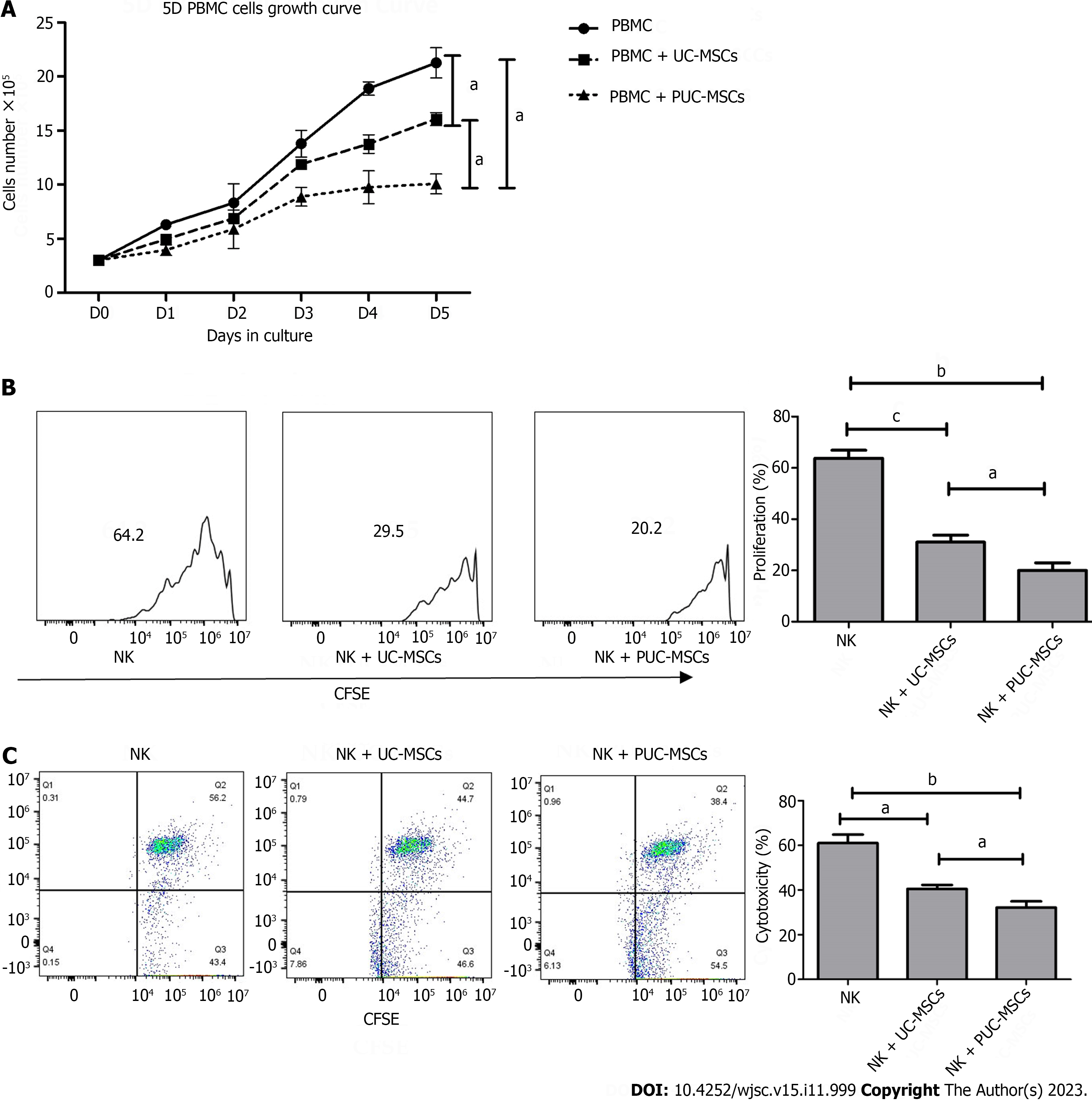
Figure 6 Detection of the immunomodulatory function of umbilical cord mesenchymal stem cells after preconditioning.
A: Peripheral blood mononuclear cells (PBMCs) alone or directly exposed to umbilical cord mesenchymal stem cells (UC-MSCs/primed UC-MSCs) for 5 d in a 3:1 ratio. The number of PBMCs in three groups was measured daily and the growth curve was plotted; B: Carboxyfluorescein diacetate succinimidyl ester (CFSE)-stained natural killer (NK) cells were cultured alone or cocultured with UC-MSCs/PUC-MSCs at a 3:1 ratio for 72 h. NK cells were harvested, and the fluorescence intensity of CFSE was measured by flow cytometry to measure the proliferation rate of NK cells; C: NK cells were collected and incubated with CFSE-stained K562 target cells for 4 h at an effector ratio (E:T = 1:5). Effector cell (NK cell)-mediated cytotoxicity of K562 cells was analyzed by flow cytometry. aP < 0.05; bP < 0.01; cP < 0.001; dP > 0.05.
- Citation: Li H, Ji XQ, Zhang SM, Bi RH. Hypoxia and inflammatory factor preconditioning enhances the immunosuppressive properties of human umbilical cord mesenchymal stem cells. World J Stem Cells 2023; 15(11): 999-1016
- URL: https://www.wjgnet.com/1948-0210/full/v15/i11/999.htm
- DOI: https://dx.doi.org/10.4252/wjsc.v15.i11.999


