Copyright
©The Author(s) 2023.
World J Stem Cells. Nov 26, 2023; 15(11): 999-1016
Published online Nov 26, 2023. doi: 10.4252/wjsc.v15.i11.999
Published online Nov 26, 2023. doi: 10.4252/wjsc.v15.i11.999
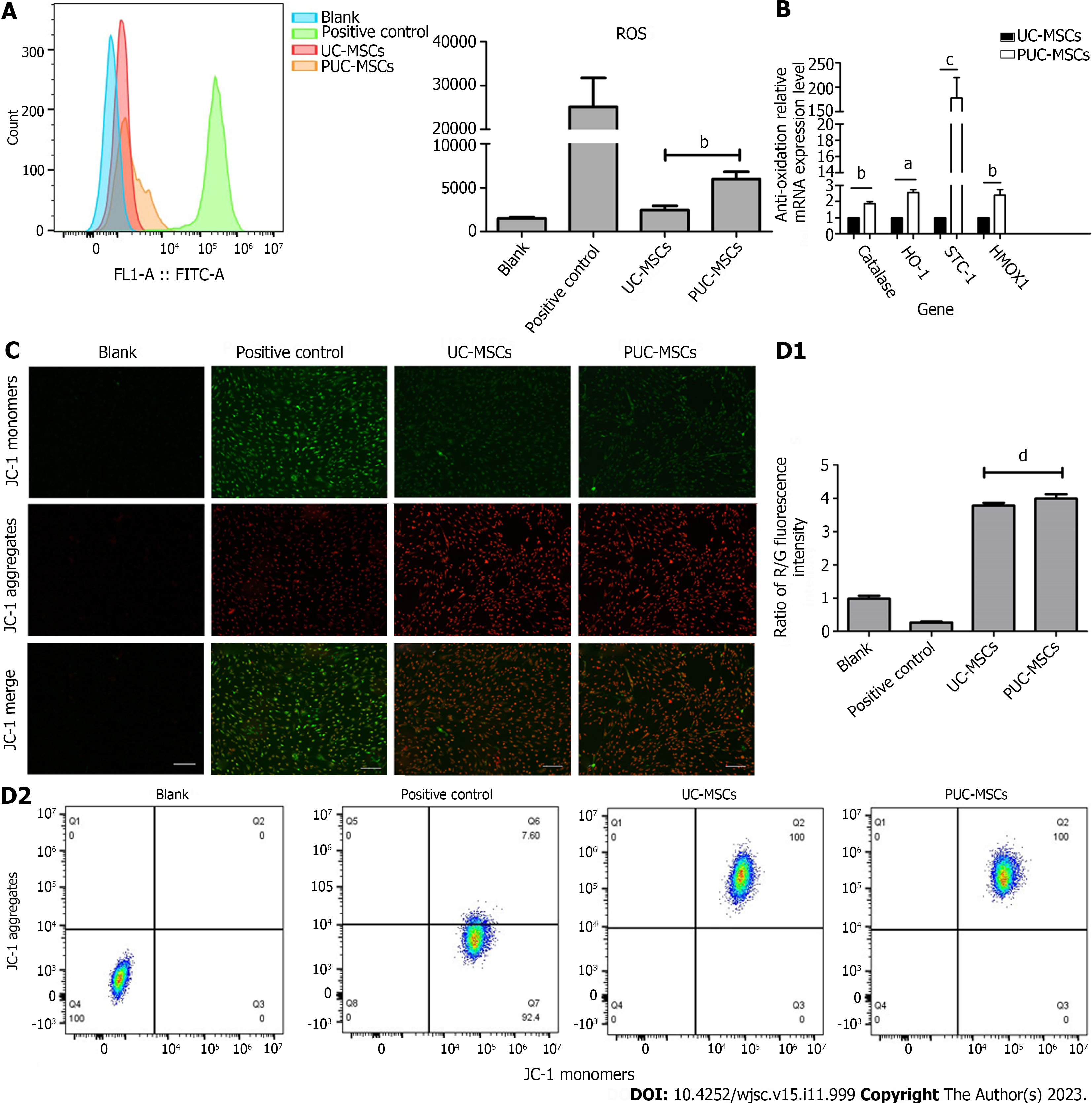
Figure 3 Mitochondrial function.
A: Reactive oxygen species (ROS) levels in umbilical cord mesenchymal stem cells (UC-MSCs) and primed UC-MSCs (PUC-MSCs) were analyzed by flow cytometry. The fluorescence intensity of 2,7-dichlorodihydrofluorescein (DCFH-DA) in PUC-MSCs increased by 3-fold compared to UC-MSCs; B: The expression of the antioxidant-related genes catalase, stanniocalcin-1 (STC1), and hemeoxygenase-1 (HOMX1) was increased; C: Representative image showing the mitochondrial membrane potential (MMP) of mesenchymal stem cells stained with JC-1. The red fluorescence of potential-dependent JC-1 aggregation in each group and the green fluorescence of the JC-1 monomer in the cytoplasm after depolarization of the mitochondrial membrane were observed. Fluorescence microscopy showed no significant difference in red and green fluorescence between UC-MSCs and PU-MSCs; Bar = 200 μm; D: Flow cytometry was used to detect the red and green fluorescence of the JC-1 probe in UC-MSCs and PUC-MSCs. There was no difference in the ratios of red and green fluorescence. aP < 0.05; bP < 0.01; cP < 0.001; dP > 0.05.
- Citation: Li H, Ji XQ, Zhang SM, Bi RH. Hypoxia and inflammatory factor preconditioning enhances the immunosuppressive properties of human umbilical cord mesenchymal stem cells. World J Stem Cells 2023; 15(11): 999-1016
- URL: https://www.wjgnet.com/1948-0210/full/v15/i11/999.htm
- DOI: https://dx.doi.org/10.4252/wjsc.v15.i11.999


