Copyright
©The Author(s) 2023.
World J Stem Cells. Nov 26, 2023; 15(11): 1017-1034
Published online Nov 26, 2023. doi: 10.4252/wjsc.v15.i11.1017
Published online Nov 26, 2023. doi: 10.4252/wjsc.v15.i11.1017
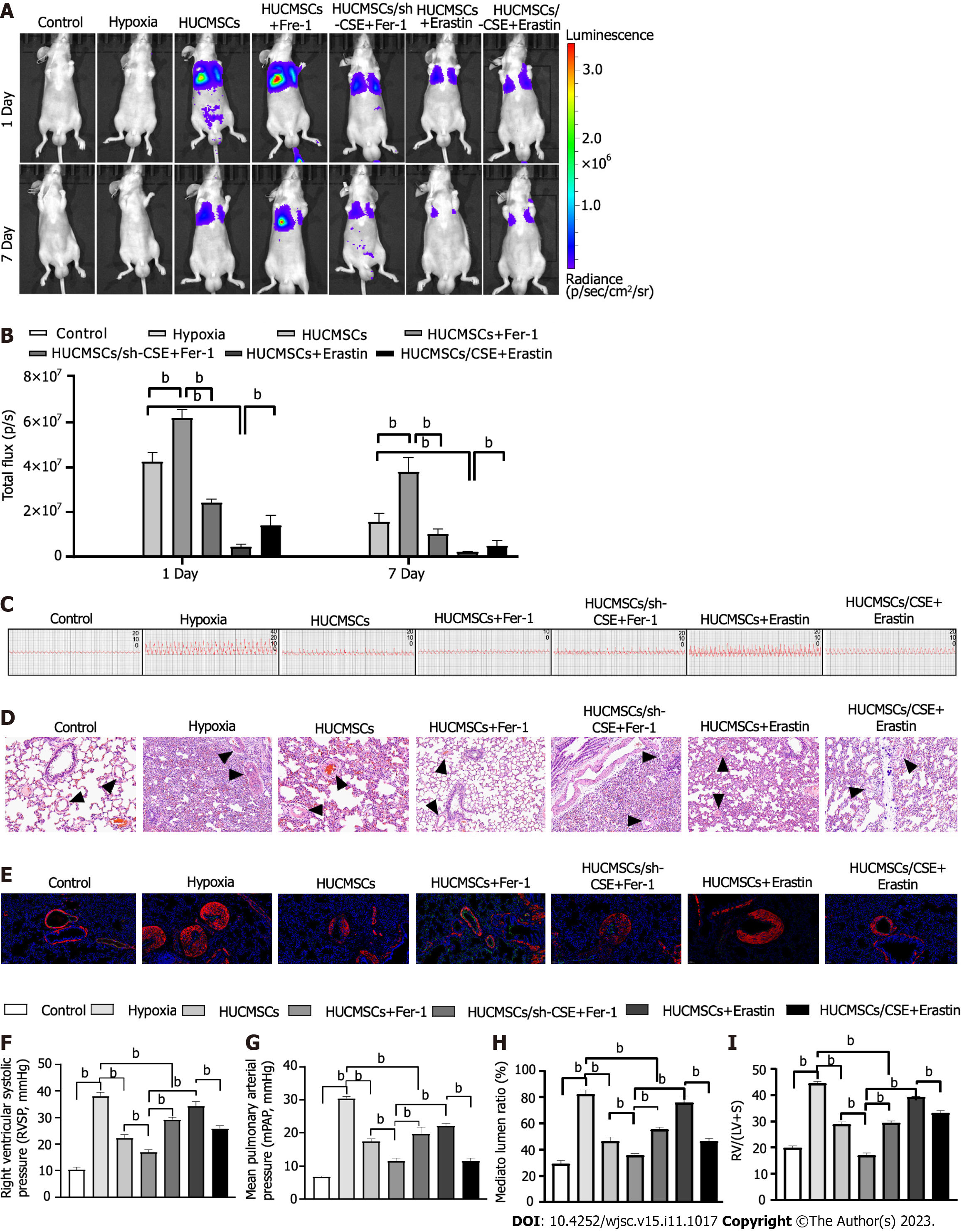
Figure 3 Change of pulmonary arterial remodeling.
A: In vivo bioluminescence imaging (BLI). Representative time course image showing in vivo BLI of mice grafted with living human umbilical cord mesenchymal stem cells expressing firefly luciferase gene in the lungs. Images were acquired at 24 h and 7 d post-implantation. Regions of interest are drawn on the mouse lung and on the mouse shoulder, considered as background signal; B: In vivo BLI. Quantitative analysis of in vivo BLI at days 1 and 7 post-implantation; C-H: Representing pulmonary arterial pressure (C, F, and G). Representative haematoxylin and eosin-stained lung sections and quantification of the ratio of the medial wall area to the total vessel cross sectional area of the distal pulmonary artery sections (D and H). Immunofluorescent staining (E). Green fluorescence represents VEcadherin, red fluorescence represents alpha-smooth muscle actin, and blue fluorescence indicates 4’,6-diamidino-2-phenylindole nuclear staining; I: Right ventricle/left ventricle plus septum. Data are expressed as the mean ± SEM (n = 6/group). HUCMSCS: Human umbilical cord mesenchymal stem cells; Fer-1: Ferrostatin-1; CSE: Cystathionine γ-lyase; RV/(LV + S): Right ventricle/(left ventricle plus septum). bP < 0.01.
- Citation: Hu B, Zhang XX, Zhang T, Yu WC. Dissecting molecular mechanisms underlying ferroptosis in human umbilical cord mesenchymal stem cells: Role of cystathionine γ-lyase/hydrogen sulfide pathway. World J Stem Cells 2023; 15(11): 1017-1034
- URL: https://www.wjgnet.com/1948-0210/full/v15/i11/1017.htm
- DOI: https://dx.doi.org/10.4252/wjsc.v15.i11.1017


