Copyright
©The Author(s) 2022.
World J Stem Cells. Sep 26, 2022; 14(9): 729-743
Published online Sep 26, 2022. doi: 10.4252/wjsc.v14.i9.729
Published online Sep 26, 2022. doi: 10.4252/wjsc.v14.i9.729
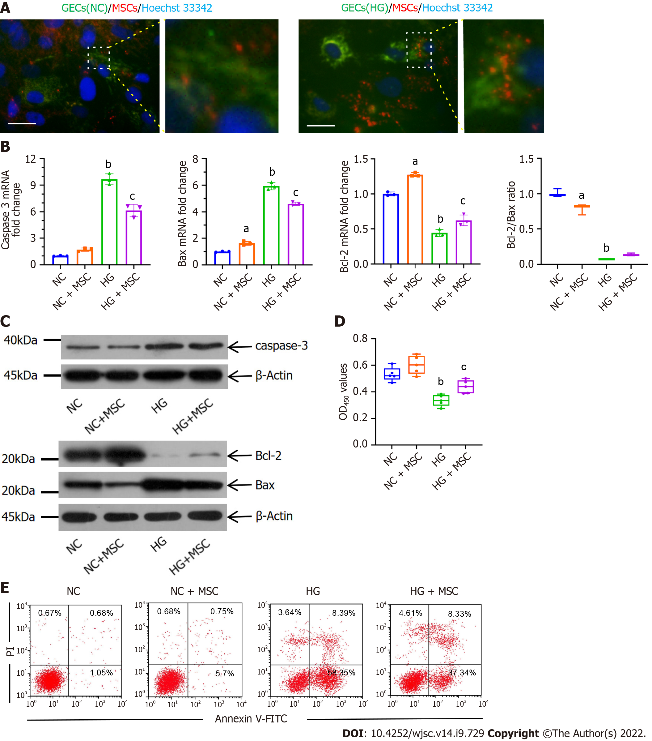
Figure 1 Anti-apoptotic effects of mesenchymal stem cells on high glucose-induced glomerular endothelial cells in vitro.
A: Immunofluorescence images of MitoTracker green (green) labeled normal control-glomerular endothelial cells (NC-GECs) and high glucose-induced GECs (HG-GECs) cultured with MitoTracker Red CMXRos (red) labeled mesenchymal stem cells (MSCs). GEC nuclei were counterstained with Hoechst 33342 (blue). A few spontaneous mitochondria transferred from MSCs to NC-GECs. Interestingly, a robust transfer of numerous mitochondria from MSCs to HG-GECs was observed. Scale bar: 200 nm; B: Caspase 3, Bax, and B-cell lymphoma 2 mRNA expression detected by real-time reverse transcriptase-polymerase chain reaction; C: Caspase 3, Bax, and B-cell lymphoma 2 protein expression detected by western blot; D: GEC viability assays performed using the Cell Counting Kit-8; E: Cellular apoptosis analysis in GEC cells treated with MSCs using Annexin V-FITC/PI staining. Data are presented as the mean ± SD. aP < 0.05 vs normal control group, bP < 0.01 vs normal control group, cP < 0.05 vs high glucose-induced group. GECs: Glomerular endothelial cells; MSCs: Mesenchymal stem cells; NC-GECs: Normal control glomerular endothelial cells; HG-GECs: High glucose-induced GECs; Bcl-2: B-cell lymphoma 2.
- Citation: Tang LX, Wei B, Jiang LY, Ying YY, Li K, Chen TX, Huang RF, Shi MJ, Xu H. Intercellular mitochondrial transfer as a means of revitalizing injured glomerular endothelial cells. World J Stem Cells 2022; 14(9): 729-743
- URL: https://www.wjgnet.com/1948-0210/full/v14/i9/729.htm
- DOI: https://dx.doi.org/10.4252/wjsc.v14.i9.729


