Copyright
©The Author(s) 2022.
World J Stem Cells. Sep 26, 2022; 14(9): 714-728
Published online Sep 26, 2022. doi: 10.4252/wjsc.v14.i9.714
Published online Sep 26, 2022. doi: 10.4252/wjsc.v14.i9.714
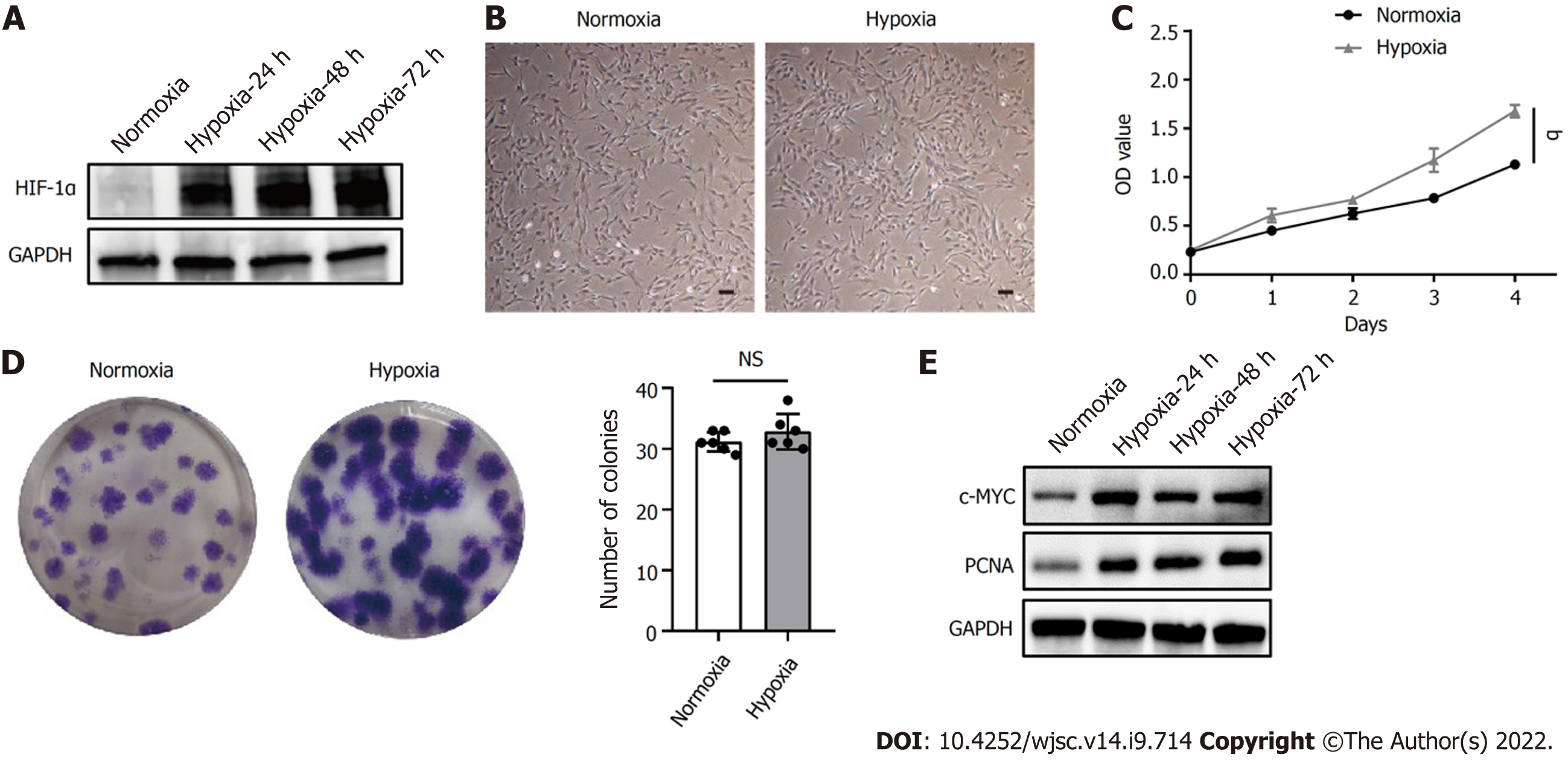
Figure 1 Hypoxia facilitated human placenta-derived mesenchymal stem cell growth and proliferation.
A: Western blot analysis of hypoxia-inducible factor 1α expression in human placenta-derived mesenchymal stem cells (hP-MSCs) under hypoxic culture for 24, 48, or 72 h; B: Morphology of the cultured hP-MSCs under hypoxia (scale bars, 100 μm); C: Proliferation curves of hP-MSCs were established based on cumulative cell numbers at different incubation times (0, 1, 2, 3, and 4 d) under normoxia or hypoxia; D: Colony size and colony number of hP-MSCs under normoxic or hypoxic culture (n = 6); E: The protein expression of c-MYC and proliferating cell nuclear antigen in hP-MSCs under hypoxic culture for 24, 48, or 72 h. Data are presented as means ± SD. aP < 0.05. NS: No significance; PCNA: Proliferating cell nuclear antigen; HIF-1α: Hypoxia-inducible factor 1α.
- Citation: Feng XD, Zhou JH, Chen JY, Feng B, Hu RT, Wu J, Pan QL, Yang JF, Yu J, Cao HC. Long non-coding RNA SNHG16 promotes human placenta-derived mesenchymal stem cell proliferation capacity through the PI3K/AKT pathway under hypoxia. World J Stem Cells 2022; 14(9): 714-728
- URL: https://www.wjgnet.com/1948-0210/full/v14/i9/714.htm
- DOI: https://dx.doi.org/10.4252/wjsc.v14.i9.714


