Copyright
©The Author(s) 2022.
World J Stem Cells. Aug 26, 2022; 14(8): 633-657
Published online Aug 26, 2022. doi: 10.4252/wjsc.v14.i8.633
Published online Aug 26, 2022. doi: 10.4252/wjsc.v14.i8.633
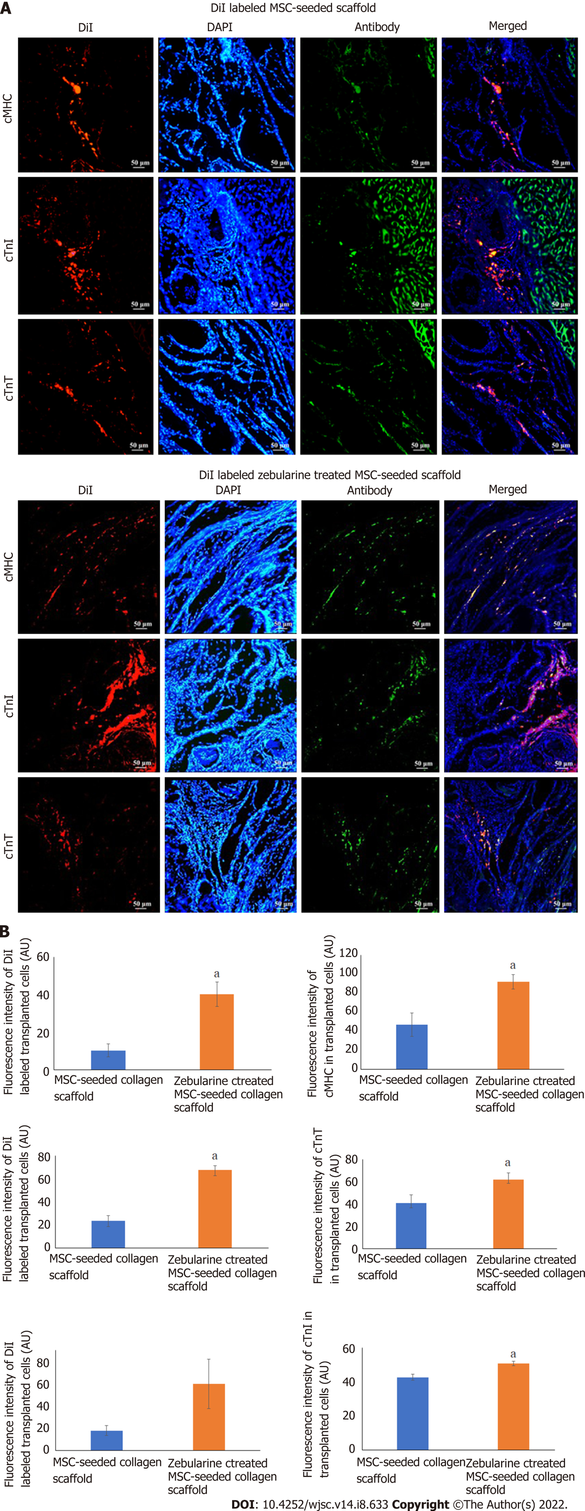
Figure 10 Cell tracking analysis.
A: Fluorescence images showing DiI labeled mesenchymal stem cells (MSCs) in the scaffold near the border of the scaffold and the infarcted myocardium, and co-localized green fluorescent cardiac proteins, cardiac myosin heavy chain, cardiac troponin T, and cardiac troponin I in the untreated and zebularine treated MSC-seeded scaffold transplanted rat heart; B: Quantification of fluorescence intensities showing significantly higher intensity of DiI labeled MSCs and protein expression in zebularine treated MSC-seeded scaffold group. Data is presented as mean ± standard error of mean with significance level aP < 0.05 (where aP < 0.05). MSC: Mesenchymal stem cells; DAPI: 4',6-diamidino-2-phenylindole. cTnT: Cardiac troponin T; cTnI: Cardiac troponin I; cMHC: Cardiac myosin heavy chain; DAPI: 4',6-diamidino-2-phenylindole.
- Citation: Qazi REM, Khan I, Haneef K, Malick TS, Naeem N, Ahmad W, Salim A, Mohsin S. Combination of mesenchymal stem cells and three-dimensional collagen scaffold preserves ventricular remodeling in rat myocardial infarction model. World J Stem Cells 2022; 14(8): 633-657
- URL: https://www.wjgnet.com/1948-0210/full/v14/i8/633.htm
- DOI: https://dx.doi.org/10.4252/wjsc.v14.i8.633


