Copyright
©The Author(s) 2022.
World J Stem Cells. Aug 26, 2022; 14(8): 633-657
Published online Aug 26, 2022. doi: 10.4252/wjsc.v14.i8.633
Published online Aug 26, 2022. doi: 10.4252/wjsc.v14.i8.633
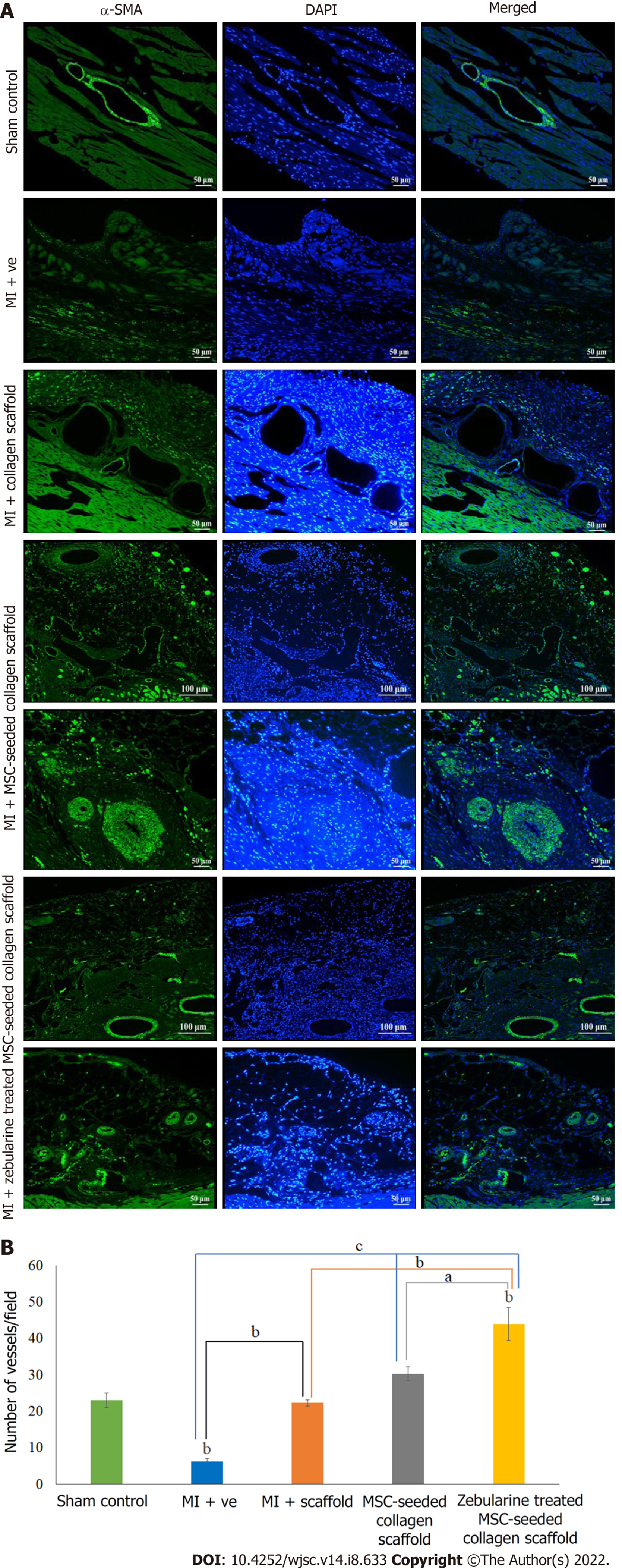
Figure 9 Analysis of angiogenesis.
A: Immunohistochemical analysis of rat hearts by α-smooth muscle actin showing blood vessels in the left ventricle in sham control and myocardial infarction model; scaffold only group, and untreated and zebularine treated mesenchymal stem cells (MSC)-seeded scaffold groups also show angiogenesis in the scaffold region as well; B: Bar graphs showing the quantification of blood vessels and statistical analysis. Zebularine treated MSC-seeded scaffold transplanted group showed more significant increase in the number of blood vessels. Data is presented as mean ± standard error of mean with significance level aP < 0.05 (where aP < 0.05, bP < 0.01, cP < 0.001). SMA: Smooth muscle actin; MI: Myocardial infarction; MSC: Mesenchymal stem cells.
- Citation: Qazi REM, Khan I, Haneef K, Malick TS, Naeem N, Ahmad W, Salim A, Mohsin S. Combination of mesenchymal stem cells and three-dimensional collagen scaffold preserves ventricular remodeling in rat myocardial infarction model. World J Stem Cells 2022; 14(8): 633-657
- URL: https://www.wjgnet.com/1948-0210/full/v14/i8/633.htm
- DOI: https://dx.doi.org/10.4252/wjsc.v14.i8.633


