Copyright
©The Author(s) 2022.
World J Stem Cells. Aug 26, 2022; 14(8): 633-657
Published online Aug 26, 2022. doi: 10.4252/wjsc.v14.i8.633
Published online Aug 26, 2022. doi: 10.4252/wjsc.v14.i8.633
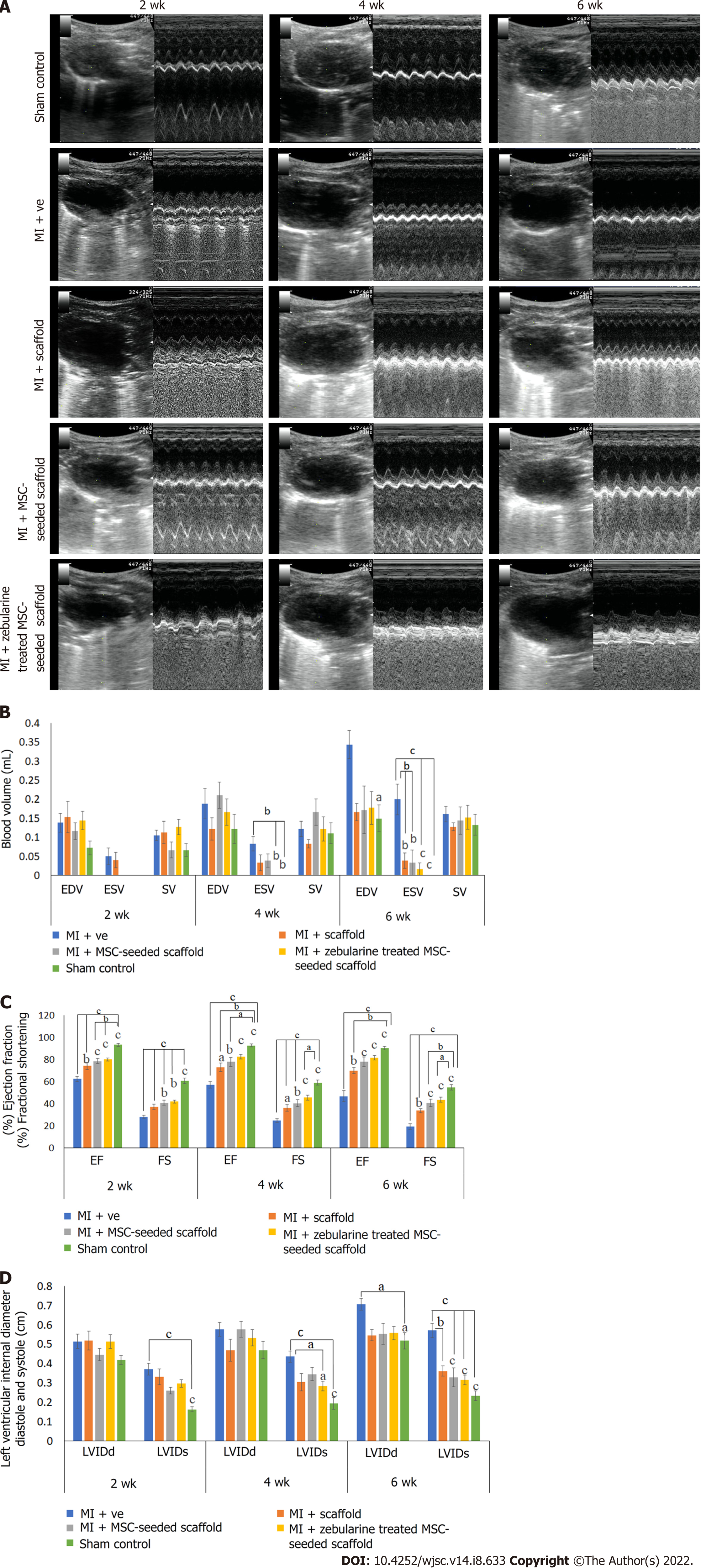
Figure 7 Cardiac functional analysis by echocardiography.
A: Ultrasound images showing B and M mode scans of left ventricle in sham control, myocardial infarction (MI) group, scaffold only group, and untreated and zebularine treated mesenchymal stem cells (MSC)-seeded scaffold groups; B-D: Bar graphs showing functional analysis in terms of (B) end systolic and diastolic volumes and stroke volume, (C) ejection fraction and fractional shortening, and (D) left ventricular systolic and diastolic internal dimensions after 2, 4 and 6 wk of MI model development. Both untreated and zebularine treated MSC-seeded collagen scaffold transplanted groups showed functional improvement. However, zebularine treated group showed more pronounced results. Data is presented as mean ± standard error of mean with significance level aP < 0.05 (where aP < 0.05, bP < 0.01, cP < 0.001). MI: Myocardial infarction; MSC: Mesenchymal stem cells; EDV: End diastolic volume; ESV: End systolic volume; LVIDd: Left ventricular internal diameter diastole; LVIDs: Left ventricular internal diameter systole; EF: Ejection fraction; FS: Fractional shortening; SV: Stroke volume.
- Citation: Qazi REM, Khan I, Haneef K, Malick TS, Naeem N, Ahmad W, Salim A, Mohsin S. Combination of mesenchymal stem cells and three-dimensional collagen scaffold preserves ventricular remodeling in rat myocardial infarction model. World J Stem Cells 2022; 14(8): 633-657
- URL: https://www.wjgnet.com/1948-0210/full/v14/i8/633.htm
- DOI: https://dx.doi.org/10.4252/wjsc.v14.i8.633


