Copyright
©The Author(s) 2022.
World J Stem Cells. Aug 26, 2022; 14(8): 616-632
Published online Aug 26, 2022. doi: 10.4252/wjsc.v14.i8.616
Published online Aug 26, 2022. doi: 10.4252/wjsc.v14.i8.616
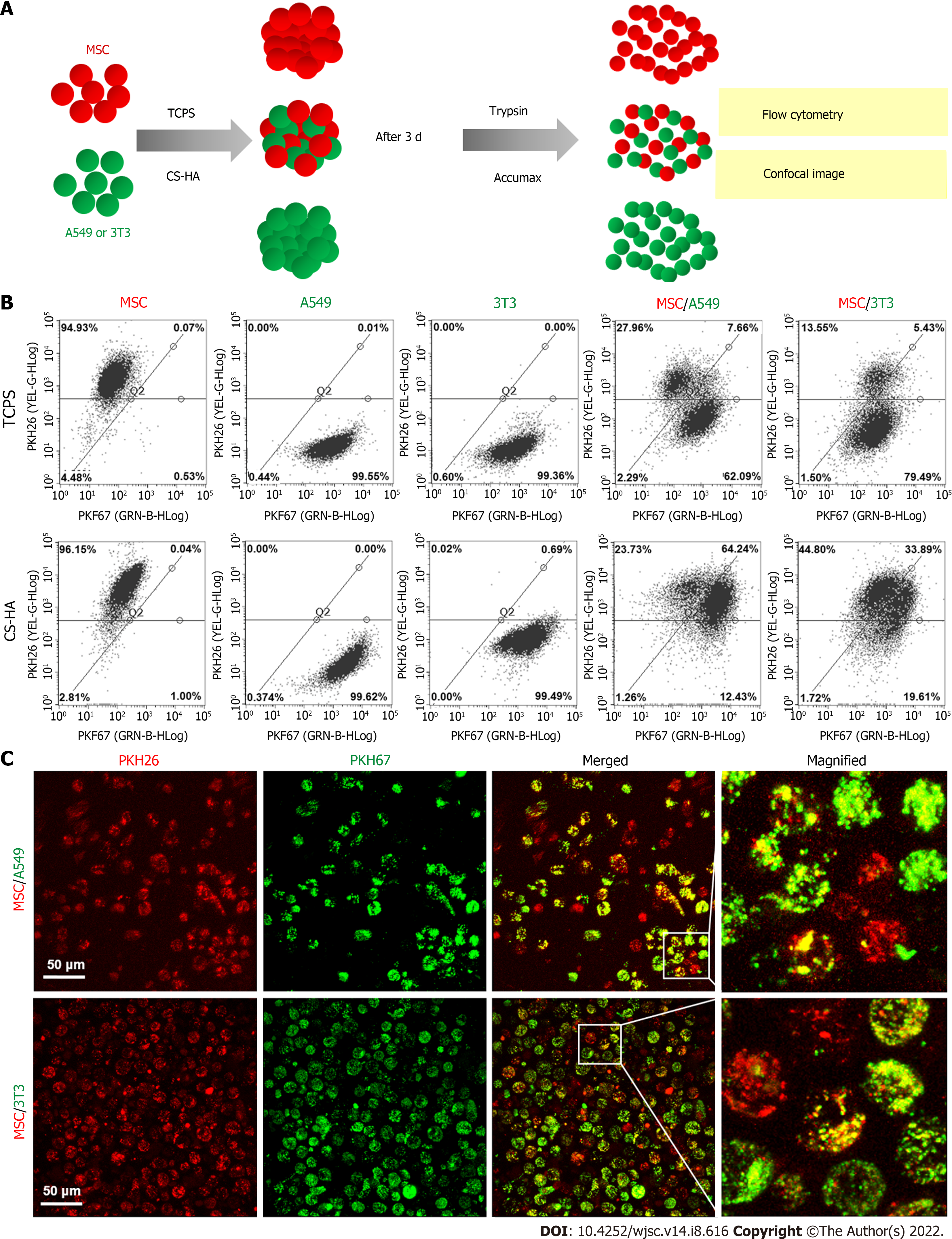
Figure 7 Membrane fluidity of cells for co-cultured cellular spheroids on tissue culture polystyrene or chitosan-hyaluronan substrates.
Membrane fluidity was determined by translocation of the membrane labeling fluorescent dyes. A: Illustration of experimental procedures for the determination of membrane fluidity. Fluorescent dye-labeled mesenchymal stem cells (MSCs) (PKH26, red), A549 (PKH67, green), or 3T3 (PKH67, green) cells were cultured on tissue culture polystyrene (TCPS) or chitosan-hyaluronan (CS-HA) substrates for 3 d. Then, the cells on TCPS were detached by trypsin and the co-spheroids on CS-HA were dissociated by Accumax. The single cell suspension was subjected to imaging and flow cytometry analysis to obtain the respective ratios of cells labeled with single and double fluorescent dyes; B: Respective ratios of the cells labeled with single and double fluorescent dyes determined by flow cytometry; C: The confocal image of single cells dissociated from MSC/A549 or MSC/3T3 co-spheroids by Accumax. Scale bar: 50 μm. TCPS: Tissue culture polystyrene; CS-HA: Chitosan-hyaluronan; MSC: Mesenchymal stem cells.
- Citation: Wong CW, Han HW, Hsu SH. Changes of cell membrane fluidity for mesenchymal stem cell spheroids on biomaterial surfaces. World J Stem Cells 2022; 14(8): 616-632
- URL: https://www.wjgnet.com/1948-0210/full/v14/i8/616.htm
- DOI: https://dx.doi.org/10.4252/wjsc.v14.i8.616


