Copyright
©The Author(s) 2022.
World J Stem Cells. Aug 26, 2022; 14(8): 616-632
Published online Aug 26, 2022. doi: 10.4252/wjsc.v14.i8.616
Published online Aug 26, 2022. doi: 10.4252/wjsc.v14.i8.616
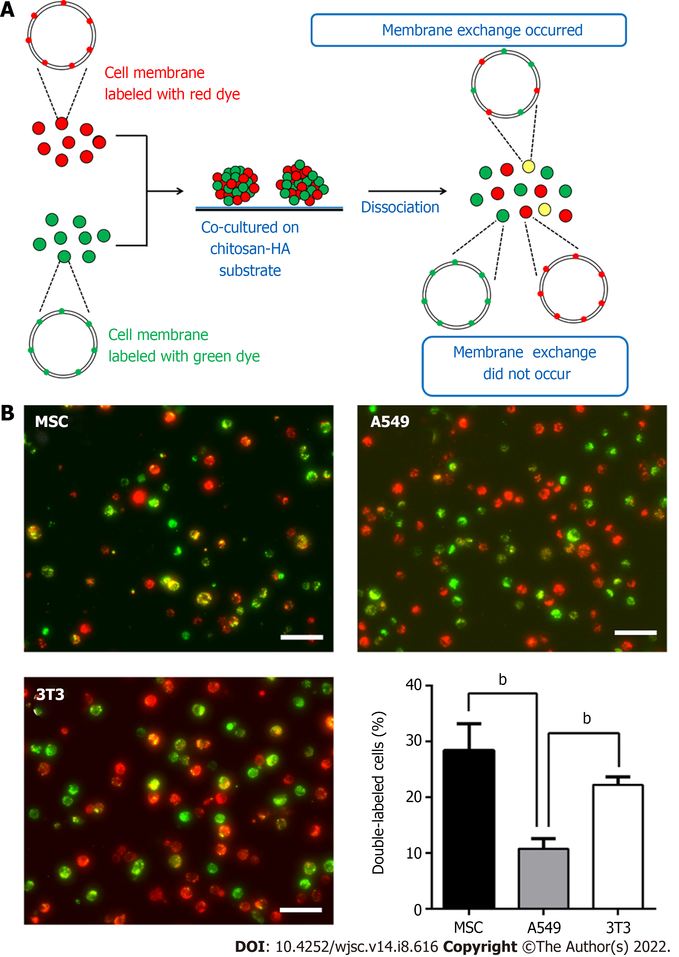
Figure 5 Membrane fluidity of mesenchymal stem cells, 3T3, and A549 individually cultured on chitosan-hyaluronan substrates.
The single cell suspension was subjected to the analysis for respective ratios of the cells labeled with single and double fluorescent dyes. A: Illustration of experimental procedures; B: Mesenchymal stem cells, A549, or 3T3 cells labeled with fluorescent dyes were cultured on chitosan-hyaluronan substrates for 3 d, and then the membrane fluidity of cells was revealed by the percentages of the double-labeled cell population. bP < 0.01, among the indicated groups. Scale bar: 100 μm. MSC: Mesenchymal stem cells.
- Citation: Wong CW, Han HW, Hsu SH. Changes of cell membrane fluidity for mesenchymal stem cell spheroids on biomaterial surfaces. World J Stem Cells 2022; 14(8): 616-632
- URL: https://www.wjgnet.com/1948-0210/full/v14/i8/616.htm
- DOI: https://dx.doi.org/10.4252/wjsc.v14.i8.616


